Back

Poster Session E - Tuesday Afternoon
Category: Interventional Endoscopy
E0458 - Role of Endoscopic Ultrasound in the Diagnosis of Extrapulmonary, Disseminated Coccidiomycosis as a Cause of Recurrent Ascites and Acute Pancreatitis
Tuesday, October 25, 2022
3:00 PM – 5:00 PM ET
Location: Crown Ballroom


Venkata Pulivarthi, MD
Creighton University School of Medicine
Phoenix, AZ
Presenting Author(s)
Venkata Pulivarthi, MD1, Aida Rezaie, MD2, Chinonso Ilo, MD2, Kayvon Sotoudeh, MD2, Hadiatou Barry, MD, MPH2, Brett Hughes, MD2, Savio Reddymasu, MD2
1Creighton University School of Medicine, Phoenix, AZ; 2Creighton University, Phoenix, AZ
Introduction: Coccidiomycosis (Valley Fever) is an endemic fungal infection in the Southwestern United States caused by Coccidiodes immitis and posadassi. Extrapulmonary involvement is very rare (approximately < 1%) and diagnosis can be challenging as the yield for culture detection is low. We present the utility of endoscopic ultrasound (EUS) with fine needle aspiration (FNA) in diagnosing a rare case of extrapulmonary coccidiomycosis with recurrent pancreatitis.
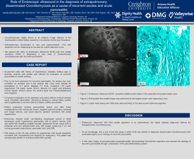
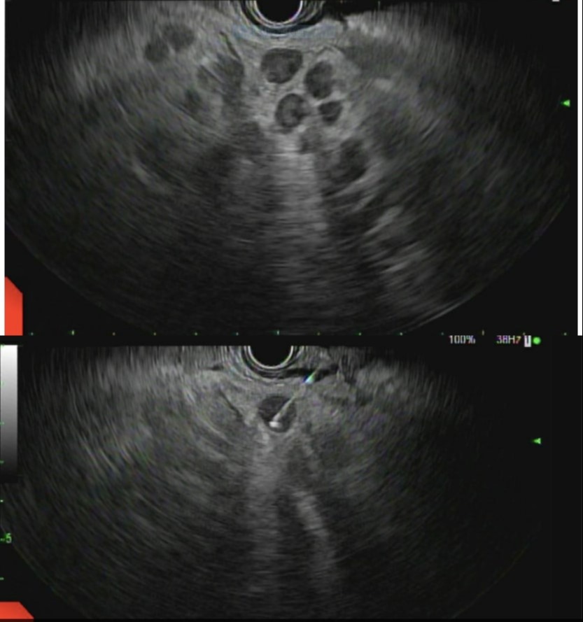
Case Description/Methods: 44 year old male with history of hypertension, diabetes mellitus type 2, alcoholic cirrhosis with ascites was referred to gastroenterology clinic for evaluation of recurrent pancreatitis of unclear etiology. Prior to his third admission for recurrent pancreatitis, his ascites was well-controlled on diuretics and a low sodium diet. Labs were notable for hemoglobin of 12.7 gm/dl, platelets 117 K/mL, creatinine 1.03 mg/dl, triglycerides 116 mg/dL, lipase 124U/L, bilirubin 2.2 mg/dL and otherwise normal hepatic function panel. His alcohol level and Phosphatidylethanol were negative. Computed tomography of the abdomen and pelvis without contrast showed acute interstitial pancreatitis. Ultrasound showed a normal biliary system without gallstones or common bile duct dilation (CBD) and ascites. Patient underwent multiple paracentesis before and after these hospitalizations with normal cell counts, negative acid-fast bacillus, bacterial and fungal cultures, and serum-albumin gradient consistent with portal hypertension. Endoscopy showed small, non-bleeding esophageal varices in distal esophagus, portal hypertensive gastropathy and no gastric varices. EUS was performed revealing multiple rounded, hypoechoic lymph nodes (LN) in periportal area and peripancreatic area with largest measuring 2 cm and normal pancreatic parenchyma, pancreatic duct, and CBD. FNA biopsy of the LN was positive for granulomas with fungal organisms consistent with Coccidioides and negative for malignancy. The patient was subsequently started on oral fluconazole 800 mg daily.
Discussion: EUS with FNA is an underutilized but highly valuable diagnostic method for extrapulmonary Coccidiomycosis. To our knowledge, this is one of the first cases in which EUS was utilized to diagnose disseminated Coccidiomycosis with lymphadenopathy as an etiology of recurrent pancreatitis. In this case, biopsy of peri-pancreatic lymphadenopathy from coccidiodes was deemed the etiology of recurrent pancreatitis by compressing pancreaticobiliary system.

Disclosures:
Venkata Pulivarthi, MD1, Aida Rezaie, MD2, Chinonso Ilo, MD2, Kayvon Sotoudeh, MD2, Hadiatou Barry, MD, MPH2, Brett Hughes, MD2, Savio Reddymasu, MD2. E0458 - Role of Endoscopic Ultrasound in the Diagnosis of Extrapulmonary, Disseminated Coccidiomycosis as a Cause of Recurrent Ascites and Acute Pancreatitis, ACG 2022 Annual Scientific Meeting Abstracts. Charlotte, NC: American College of Gastroenterology.
1Creighton University School of Medicine, Phoenix, AZ; 2Creighton University, Phoenix, AZ
Introduction: Coccidiomycosis (Valley Fever) is an endemic fungal infection in the Southwestern United States caused by Coccidiodes immitis and posadassi. Extrapulmonary involvement is very rare (approximately < 1%) and diagnosis can be challenging as the yield for culture detection is low. We present the utility of endoscopic ultrasound (EUS) with fine needle aspiration (FNA) in diagnosing a rare case of extrapulmonary coccidiomycosis with recurrent pancreatitis.
Case Description/Methods: 44 year old male with history of hypertension, diabetes mellitus type 2, alcoholic cirrhosis with ascites was referred to gastroenterology clinic for evaluation of recurrent pancreatitis of unclear etiology. Prior to his third admission for recurrent pancreatitis, his ascites was well-controlled on diuretics and a low sodium diet. Labs were notable for hemoglobin of 12.7 gm/dl, platelets 117 K/mL, creatinine 1.03 mg/dl, triglycerides 116 mg/dL, lipase 124U/L, bilirubin 2.2 mg/dL and otherwise normal hepatic function panel. His alcohol level and Phosphatidylethanol were negative. Computed tomography of the abdomen and pelvis without contrast showed acute interstitial pancreatitis. Ultrasound showed a normal biliary system without gallstones or common bile duct dilation (CBD) and ascites. Patient underwent multiple paracentesis before and after these hospitalizations with normal cell counts, negative acid-fast bacillus, bacterial and fungal cultures, and serum-albumin gradient consistent with portal hypertension. Endoscopy showed small, non-bleeding esophageal varices in distal esophagus, portal hypertensive gastropathy and no gastric varices. EUS was performed revealing multiple rounded, hypoechoic lymph nodes (LN) in periportal area and peripancreatic area with largest measuring 2 cm and normal pancreatic parenchyma, pancreatic duct, and CBD. FNA biopsy of the LN was positive for granulomas with fungal organisms consistent with Coccidioides and negative for malignancy. The patient was subsequently started on oral fluconazole 800 mg daily.
Discussion: EUS with FNA is an underutilized but highly valuable diagnostic method for extrapulmonary Coccidiomycosis. To our knowledge, this is one of the first cases in which EUS was utilized to diagnose disseminated Coccidiomycosis with lymphadenopathy as an etiology of recurrent pancreatitis. In this case, biopsy of peri-pancreatic lymphadenopathy from coccidiodes was deemed the etiology of recurrent pancreatitis by compressing pancreaticobiliary system.

Figure: Figure 1: EUS revealed multiple lymph nodes in the periportal and peripancreatic area.
Figure 2: EUS-guided fine needle biopsy was performed on the largest lymph node measuring 2 cms.
Figure 2: EUS-guided fine needle biopsy was performed on the largest lymph node measuring 2 cms.
Disclosures:
Venkata Pulivarthi indicated no relevant financial relationships.
Aida Rezaie indicated no relevant financial relationships.
Chinonso Ilo indicated no relevant financial relationships.
Kayvon Sotoudeh indicated no relevant financial relationships.
Hadiatou Barry indicated no relevant financial relationships.
Brett Hughes indicated no relevant financial relationships.
Savio Reddymasu indicated no relevant financial relationships.
Venkata Pulivarthi, MD1, Aida Rezaie, MD2, Chinonso Ilo, MD2, Kayvon Sotoudeh, MD2, Hadiatou Barry, MD, MPH2, Brett Hughes, MD2, Savio Reddymasu, MD2. E0458 - Role of Endoscopic Ultrasound in the Diagnosis of Extrapulmonary, Disseminated Coccidiomycosis as a Cause of Recurrent Ascites and Acute Pancreatitis, ACG 2022 Annual Scientific Meeting Abstracts. Charlotte, NC: American College of Gastroenterology.
