Back


Poster Session D - Tuesday Morning
Category: Colon
D0138 - Colonic Varices as a Complication of Pancreatic Adenocarcinoma: A Case Report and Literature Review
Tuesday, October 25, 2022
10:00 AM – 12:00 PM ET
Location: Crown Ballroom

Has Audio

Hima Veeramachaneni, MD
Emory University School of Medicine
Atlanta, GA
Presenting Author(s)
Hima Veeramachaneni, MD1, Abubaker Abdalla, MD1, Kiara Smith, MD1, Sanskriti Sharma, MD2, Vaishali Patel, MD1
1Emory University School of Medicine, Atlanta, GA; 2WellStar Atlanta Medical Center, Atlanta, GA
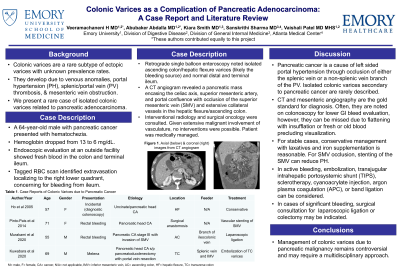
Introduction: Colonic varices are a rare subtype of ectopic varices with unknown prevalence rates, likely due to under-diagnosis. They form due to venous anomalies, portal hypertension, splenic or portal vein (PV) thrombosis and mesenteric vein obstruction. We present a rare case of isolated colonic varices related to pancreatic cancer.
Case Description/Methods: A 64-year-old male with pancreatic cancer presented with hematochezia. Endoscopic evaluation at an outside facility showed fresh blood in the colon and terminal ileum. His hemoglobin dropped from 13 to 6 mg/dL and a tagged RBC scan identified extravasation in the right lower quadrant, localizing to the ileum. He was transferred to our hospital for a retrograde single balloon enteroscopy, which noted isolated ascending colon and hepatic flexure varices (felt to be the source of bleeding) and normal ileum. A CT angiogram revealed the pancreas mass encased the celiac axis, superior mesenteric artery, and portal confluence with occlusion of the superior mesenteric vein (SMV) and extensive collateral vessels in the hepatic flexure/ascending colon. Interventional radiology and surgical oncology were consulted for management. Given extensive vasculature involvement, no interventions were possible, and patient was medically managed.
Discussion: Pancreatic cancer is a cause of left sided portal hypertension through occlusion of either the splenic vein or a non-splenic vein branch of the PV. Isolated colonic varices secondary to pancreatic cancer are rarely described. The gold standard for diagnosis is CT or selective mesenteric angiography. Often, they are diagnosed by colonoscopy for lower GI bleed evaluation but can be missed due to flattening with insufflation or blood precluding visualization.
Treatment options are limited and mostly based on recommendations from case reports as there are no guidelines. For uncomplicated cases, conservative management with laxatives and iron supplementation is reasonable. For SMV occlusion, stenting of the SMV can reduce PH (table 1). In active bleeding, options include embolization, endoscopic therapy (sclerotherapy, cyanoacrylate injection, argon plasma coagulation, and band ligation), or transjugular intrahepatic portosystemic shunt (TIPS). However, if significant bleeding, laparoscopic ligation or colectomy may be indicated. Management of colonic varices due to pancreatic malignancy remains controversial and may require a multidisciplinary approach.
Disclosures:
Hima Veeramachaneni, MD1, Abubaker Abdalla, MD1, Kiara Smith, MD1, Sanskriti Sharma, MD2, Vaishali Patel, MD1. D0138 - Colonic Varices as a Complication of Pancreatic Adenocarcinoma: A Case Report and Literature Review, ACG 2022 Annual Scientific Meeting Abstracts. Charlotte, NC: American College of Gastroenterology.
1Emory University School of Medicine, Atlanta, GA; 2WellStar Atlanta Medical Center, Atlanta, GA
Introduction: Colonic varices are a rare subtype of ectopic varices with unknown prevalence rates, likely due to under-diagnosis. They form due to venous anomalies, portal hypertension, splenic or portal vein (PV) thrombosis and mesenteric vein obstruction. We present a rare case of isolated colonic varices related to pancreatic cancer.
Case Description/Methods: A 64-year-old male with pancreatic cancer presented with hematochezia. Endoscopic evaluation at an outside facility showed fresh blood in the colon and terminal ileum. His hemoglobin dropped from 13 to 6 mg/dL and a tagged RBC scan identified extravasation in the right lower quadrant, localizing to the ileum. He was transferred to our hospital for a retrograde single balloon enteroscopy, which noted isolated ascending colon and hepatic flexure varices (felt to be the source of bleeding) and normal ileum. A CT angiogram revealed the pancreas mass encased the celiac axis, superior mesenteric artery, and portal confluence with occlusion of the superior mesenteric vein (SMV) and extensive collateral vessels in the hepatic flexure/ascending colon. Interventional radiology and surgical oncology were consulted for management. Given extensive vasculature involvement, no interventions were possible, and patient was medically managed.
Discussion: Pancreatic cancer is a cause of left sided portal hypertension through occlusion of either the splenic vein or a non-splenic vein branch of the PV. Isolated colonic varices secondary to pancreatic cancer are rarely described. The gold standard for diagnosis is CT or selective mesenteric angiography. Often, they are diagnosed by colonoscopy for lower GI bleed evaluation but can be missed due to flattening with insufflation or blood precluding visualization.
Treatment options are limited and mostly based on recommendations from case reports as there are no guidelines. For uncomplicated cases, conservative management with laxatives and iron supplementation is reasonable. For SMV occlusion, stenting of the SMV can reduce PH (table 1). In active bleeding, options include embolization, endoscopic therapy (sclerotherapy, cyanoacrylate injection, argon plasma coagulation, and band ligation), or transjugular intrahepatic portosystemic shunt (TIPS). However, if significant bleeding, laparoscopic ligation or colectomy may be indicated. Management of colonic varices due to pancreatic malignancy remains controversial and may require a multidisciplinary approach.
Disclosures:
Hima Veeramachaneni indicated no relevant financial relationships.
Abubaker Abdalla indicated no relevant financial relationships.
Kiara Smith indicated no relevant financial relationships.
Sanskriti Sharma indicated no relevant financial relationships.
Vaishali Patel indicated no relevant financial relationships.
Hima Veeramachaneni, MD1, Abubaker Abdalla, MD1, Kiara Smith, MD1, Sanskriti Sharma, MD2, Vaishali Patel, MD1. D0138 - Colonic Varices as a Complication of Pancreatic Adenocarcinoma: A Case Report and Literature Review, ACG 2022 Annual Scientific Meeting Abstracts. Charlotte, NC: American College of Gastroenterology.
