Back


Poster Session E - Tuesday Afternoon
Category: Liver
E0546 - Metastatic Hepatic Epithelioid Hemangioendothelioma: A Case Report
Tuesday, October 25, 2022
3:00 PM – 5:00 PM ET
Location: Crown Ballroom

Has Audio

Cameron Palmer, MS
EVMS
Norfolk, VA
Presenting Author(s)
Cameron Palmer, MS1, Daniel Neumann, MD2
1EVMS, Norfolk, VA; 2EVMS, Suffolk, VA
Introduction: Epithelioid Hemangioendothelioma (EHE) is a very rare vascular neoplasm consisting of epithelioid or histiocytoid cells that have endothelial characteristics. EHE has an incidence of 0.038/100,000/year peaking in the 4th-5th decade and a prevalence of < 1/1,000,000 with a slight predominance in females. EHE can arise anywhere throughout the body but mostly involves the liver, lungs, and bone with >50% of patients presenting with metastatic disease. The variability of its nature and clinical course makes uniform staging, treatment, and prognosis difficult. Several possible causative factors have been recommended; however, they remain as loose associations or still potentially undiscovered.
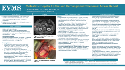
Case Description/Methods: We present a noteworthy case of this rare tumor in a previously healthy 47-year-old male, non-smoker, 1-2 EtOH 2-3 nights/wk, family history of skin cancer, UC, and lung cancer, that presented with 2 weeks of nonspecific symptoms. Initial presentation was concerning for cholecystitis; therefore, an US and CT were performed finding a complex lesion in the right hepatic lobe measuring 6.5X5.3cm. CT guided core needle bx was significant for atypical cells seen within a somewhat loose myxoid background, with occasional cytoplasmic vacuoles. Immunohistochemistry revealed (+) CD31/34 and a strong (+) CAMTA-1 nuclear staining which confirmed EHE. Additional CT chest showed several small bilateral pulmonary nodules, the largest measuring 1.1cm in the left upper lobe concerning for mets. He underwent a right total hepatectomy with associated cholecystectomy and a wedge bx of a suspicious lesion in the right lateral abdominal wall parietal peritoneum overlying the diaphragm.
Discussion: Given diagnosis of hepatic EHE with stage IV mets, Oncology planned for CT chest/abd/pelvis as part of standard surveillance with additional imaging only as indicated for symptoms not attributable to other causes. Given his small and indeterminate pulmonary nodules, close observation was favored with follow-up and repeat imaging q3mX1yr then q4mX1yr then q6mo presuming stable disease. With no standard of care for EHE, chemotherapy typically uses drugs for other soft tissue sarcomas or anti-angiogenic approaches and would be important if he has disease progression. 15 months post-op imaging showed unchanged bilateral pulmonary nodules, with stable to slightly small seroma, unchanged left renal lesions, unchanged omental infiltration, and nodularity particular in the right upper quadrant suspicious for sarcomatosis
Disclosures:
Cameron Palmer, MS1, Daniel Neumann, MD2. E0546 - Metastatic Hepatic Epithelioid Hemangioendothelioma: A Case Report, ACG 2022 Annual Scientific Meeting Abstracts. Charlotte, NC: American College of Gastroenterology.
1EVMS, Norfolk, VA; 2EVMS, Suffolk, VA
Introduction: Epithelioid Hemangioendothelioma (EHE) is a very rare vascular neoplasm consisting of epithelioid or histiocytoid cells that have endothelial characteristics. EHE has an incidence of 0.038/100,000/year peaking in the 4th-5th decade and a prevalence of < 1/1,000,000 with a slight predominance in females. EHE can arise anywhere throughout the body but mostly involves the liver, lungs, and bone with >50% of patients presenting with metastatic disease. The variability of its nature and clinical course makes uniform staging, treatment, and prognosis difficult. Several possible causative factors have been recommended; however, they remain as loose associations or still potentially undiscovered.
Case Description/Methods: We present a noteworthy case of this rare tumor in a previously healthy 47-year-old male, non-smoker, 1-2 EtOH 2-3 nights/wk, family history of skin cancer, UC, and lung cancer, that presented with 2 weeks of nonspecific symptoms. Initial presentation was concerning for cholecystitis; therefore, an US and CT were performed finding a complex lesion in the right hepatic lobe measuring 6.5X5.3cm. CT guided core needle bx was significant for atypical cells seen within a somewhat loose myxoid background, with occasional cytoplasmic vacuoles. Immunohistochemistry revealed (+) CD31/34 and a strong (+) CAMTA-1 nuclear staining which confirmed EHE. Additional CT chest showed several small bilateral pulmonary nodules, the largest measuring 1.1cm in the left upper lobe concerning for mets. He underwent a right total hepatectomy with associated cholecystectomy and a wedge bx of a suspicious lesion in the right lateral abdominal wall parietal peritoneum overlying the diaphragm.
Discussion: Given diagnosis of hepatic EHE with stage IV mets, Oncology planned for CT chest/abd/pelvis as part of standard surveillance with additional imaging only as indicated for symptoms not attributable to other causes. Given his small and indeterminate pulmonary nodules, close observation was favored with follow-up and repeat imaging q3mX1yr then q4mX1yr then q6mo presuming stable disease. With no standard of care for EHE, chemotherapy typically uses drugs for other soft tissue sarcomas or anti-angiogenic approaches and would be important if he has disease progression. 15 months post-op imaging showed unchanged bilateral pulmonary nodules, with stable to slightly small seroma, unchanged left renal lesions, unchanged omental infiltration, and nodularity particular in the right upper quadrant suspicious for sarcomatosis
Disclosures:
Cameron Palmer indicated no relevant financial relationships.
Daniel Neumann: Exact Sciences – Advisor or Review Panel Member. Optum/SCA – Advisory Committee/Board Member.
Cameron Palmer, MS1, Daniel Neumann, MD2. E0546 - Metastatic Hepatic Epithelioid Hemangioendothelioma: A Case Report, ACG 2022 Annual Scientific Meeting Abstracts. Charlotte, NC: American College of Gastroenterology.
