Back

Poster Session D - Tuesday Morning
Category: Small Intestine
D0668 - Duodenal Polyposis: An Incidental Finding of Duodenal-Type Follicular Lymphoma
Tuesday, October 25, 2022
10:00 AM – 12:00 PM ET
Location: Crown Ballroom

- AS
Anjiya Shaikh, MBBS
University of Connecticut
Hartford, CT, CT
Presenting Author(s)
Anjiya Shaikh, MBBS1, Emily Weng, DO2, Steven A. Goldenberg, MD3
1University of Connecticut, Hartford, CT; 2University of Connecticut, Farmington, CT; 3UConn Health, Farmington, CT
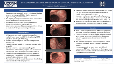
Introduction: Duodenal polyps commonly present as incidental findings on upper endoscopies (EGD), and often represent regenerative/hyperplastic nodules. The majority of neoplastic lesions are either adenomatous lesions of intestinal or gastric phenotype. A newly recognized entity of duodenal polyposis is duodenal-type follicular lymphoma (DFL). We present a rare case of duodenal polyposis from DFL as an incidental finding on a screening endoscopy
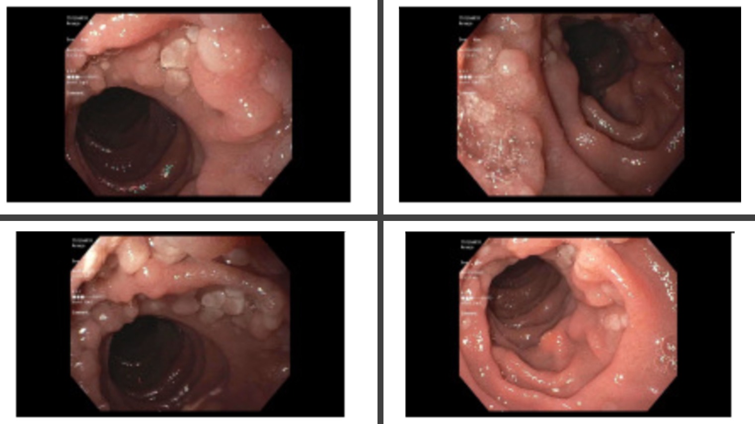
Case Description/Methods: A 58-year-old non-smoking man with no significant medical history underwent EGD for longstanding GERD symptoms refractory to PPI therapy in absence of other symptoms and unrevealing physical examination. Family history was notable for gastric carcinoma in father at age 58. Due to his risk factors, EGD was performed and notable for gastric inflammation and duodenal polyposis with multiple semi-sessile polyps in the second portion of the duodenum. Gastric biopsies revealed moderate acute and chronic gastritis, with a positive immunohistochemical stain for H-Pylori. Duodenal mucosa had abundant lymphoid aggregates, comprising of mainly CD20+ B-cells. B-cell follicles were positive for BCL2, BCL6, and CD10. Alongside the markedly nodular duodenum, these findings were consistent with B-cell lymphoma.
Laboratory studies were largely unremarkable with normal CBC with borderline high uric acid 8.0 mg/dl (3.4-7.8 mg/dl), and LDH 226 u/L (125-220). Secondary involvement by a systemic B-cell lymphoma was excluded by a normal bone marrow and PET scan. Whole body PET scan was negative for any evidence of FDG avid malignancy. Patient was diagnosed with a lowgrade, stage-1 FL. Treatment options included localized radiotherapy with a curative intent or expectant monitoring off therapy. The patient’s chronic GERD symptoms were attributed to gastritis caused by H. pylori, which isn’t considered to play a role in the development of DFL.
Discussion: DFL is a newly recognized variant of follicular lymphoma, with a low grade on presentation and benign evolution. The most common endoscopic finding is the presence of white nodular lesions confined within the duodenum, as described in this case, with rare appearances in other areas of the small intestine. Due to excellent prognosis and rare progression even in absence of treatment, watchful waiting is frequently favored. Endoscopists should be aware of the well-defined appearance of DFL, and the importance of ruling out extra duodenal involvement in order to present the patient with appropriate treatment options.

Disclosures:
Anjiya Shaikh, MBBS1, Emily Weng, DO2, Steven A. Goldenberg, MD3. D0668 - Duodenal Polyposis: An Incidental Finding of Duodenal-Type Follicular Lymphoma, ACG 2022 Annual Scientific Meeting Abstracts. Charlotte, NC: American College of Gastroenterology.
1University of Connecticut, Hartford, CT; 2University of Connecticut, Farmington, CT; 3UConn Health, Farmington, CT
Introduction: Duodenal polyps commonly present as incidental findings on upper endoscopies (EGD), and often represent regenerative/hyperplastic nodules. The majority of neoplastic lesions are either adenomatous lesions of intestinal or gastric phenotype. A newly recognized entity of duodenal polyposis is duodenal-type follicular lymphoma (DFL). We present a rare case of duodenal polyposis from DFL as an incidental finding on a screening endoscopy
Case Description/Methods: A 58-year-old non-smoking man with no significant medical history underwent EGD for longstanding GERD symptoms refractory to PPI therapy in absence of other symptoms and unrevealing physical examination. Family history was notable for gastric carcinoma in father at age 58. Due to his risk factors, EGD was performed and notable for gastric inflammation and duodenal polyposis with multiple semi-sessile polyps in the second portion of the duodenum. Gastric biopsies revealed moderate acute and chronic gastritis, with a positive immunohistochemical stain for H-Pylori. Duodenal mucosa had abundant lymphoid aggregates, comprising of mainly CD20+ B-cells. B-cell follicles were positive for BCL2, BCL6, and CD10. Alongside the markedly nodular duodenum, these findings were consistent with B-cell lymphoma.
Laboratory studies were largely unremarkable with normal CBC with borderline high uric acid 8.0 mg/dl (3.4-7.8 mg/dl), and LDH 226 u/L (125-220). Secondary involvement by a systemic B-cell lymphoma was excluded by a normal bone marrow and PET scan. Whole body PET scan was negative for any evidence of FDG avid malignancy. Patient was diagnosed with a lowgrade, stage-1 FL. Treatment options included localized radiotherapy with a curative intent or expectant monitoring off therapy. The patient’s chronic GERD symptoms were attributed to gastritis caused by H. pylori, which isn’t considered to play a role in the development of DFL.
Discussion: DFL is a newly recognized variant of follicular lymphoma, with a low grade on presentation and benign evolution. The most common endoscopic finding is the presence of white nodular lesions confined within the duodenum, as described in this case, with rare appearances in other areas of the small intestine. Due to excellent prognosis and rare progression even in absence of treatment, watchful waiting is frequently favored. Endoscopists should be aware of the well-defined appearance of DFL, and the importance of ruling out extra duodenal involvement in order to present the patient with appropriate treatment options.

Figure: Polyposis noted in the second portion of the Duodenum
Disclosures:
Anjiya Shaikh indicated no relevant financial relationships.
Emily Weng indicated no relevant financial relationships.
Steven Goldenberg indicated no relevant financial relationships.
Anjiya Shaikh, MBBS1, Emily Weng, DO2, Steven A. Goldenberg, MD3. D0668 - Duodenal Polyposis: An Incidental Finding of Duodenal-Type Follicular Lymphoma, ACG 2022 Annual Scientific Meeting Abstracts. Charlotte, NC: American College of Gastroenterology.
