Back


Poster Session E - Tuesday Afternoon
Category: Stomach
E0730 - Choroidal Melanoma: A Rare Cause of Gastric/Duodenal Pigmented Macules
Tuesday, October 25, 2022
3:00 PM – 5:00 PM ET
Location: Crown Ballroom

Has Audio

Romy Chamoun, MD
Lankenau Medical Center
Wynnewood, PA
Presenting Author(s)
Romy Chamoun, MD1, Jared Lander, DO2, Jack A. Collazzo, MD3
1Lankenau Medical Center, Wynnewood, PA; 2Lankenau Medical Center, Havertown, PA; 3Bryn Mawr Medical Specialists Association, Bryn Mawr, PA
Introduction: Choroidal melanoma is the most common primary intraocular malignancy. Two to four percent of newly diagnosed patients have evidence of metastatic disease. Up to half of patients will develop metastatic disease. The liver is the primary site of metastasis as the tumor spreads hematogenously. Metastasis to the gastrointestinal tract is rare in ocular melanoma in contrast to cutaneous melanoma. We present a case of ocular melanoma with metastasis to the gastrointestinal (GI) tract.
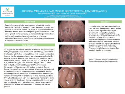
Case Description/Methods: An 81-year-old female with a history of choroidal melanoma of the left eye diagnosed in 2015 treated with radiotherapy presented to the hospital with fatigue and weight loss of 30 pounds over the last few months. On admission she was normotensive and afebrile. On physical exam her abdomen was distended and nontender. Labs were notable for Cr 2.3 mg/dL, AST 698 IU/L, ALT 286 IU/L, ALP 488 IU/L, Albumin 2.3 g/dL, Total bilirubin 4.9 mg/dL, WBC 13.3 K/uL, Hgb 11.7 g/dL, platelets 206 K/uL and INR 1.6. Computed tomography without contrast of the abdomen/pelvis showed lobular contour of the liver with multiple hyperdense foci scattered throughout concerning for metastases. Ultrasound with dopplers revealed portal vein thrombosis. Patient underwent endoscopy for variceal screening with no evidence of varices. However, scattered throughout the stomach were hyperpigmented macules of variable size (a, b). In her duodenum, there were additional lesions (c) and two non-bleeding ulcers with pigmented lesions. Biopsies were consistent with metastatic melanoma. Ultimately, hospice was pursued.
Discussion: Choroidal melanoma metastases in the GI tract are rare. When patients with a history of melanoma, regardless of its origin, present with nonspecific symptoms, physicians should have a high suspicion for metastatic disease. Metastases are endoscopically diagnosed as pigmented or amelanotic nodules, ulcerations, macules or mass. Patients are typically treated with palliative surgery or immunotherapy. Prognosis is typically poor with a median survival rate of 4 to 6 months.

Disclosures:
Romy Chamoun, MD1, Jared Lander, DO2, Jack A. Collazzo, MD3. E0730 - Choroidal Melanoma: A Rare Cause of Gastric/Duodenal Pigmented Macules, ACG 2022 Annual Scientific Meeting Abstracts. Charlotte, NC: American College of Gastroenterology.
1Lankenau Medical Center, Wynnewood, PA; 2Lankenau Medical Center, Havertown, PA; 3Bryn Mawr Medical Specialists Association, Bryn Mawr, PA
Introduction: Choroidal melanoma is the most common primary intraocular malignancy. Two to four percent of newly diagnosed patients have evidence of metastatic disease. Up to half of patients will develop metastatic disease. The liver is the primary site of metastasis as the tumor spreads hematogenously. Metastasis to the gastrointestinal tract is rare in ocular melanoma in contrast to cutaneous melanoma. We present a case of ocular melanoma with metastasis to the gastrointestinal (GI) tract.
Case Description/Methods: An 81-year-old female with a history of choroidal melanoma of the left eye diagnosed in 2015 treated with radiotherapy presented to the hospital with fatigue and weight loss of 30 pounds over the last few months. On admission she was normotensive and afebrile. On physical exam her abdomen was distended and nontender. Labs were notable for Cr 2.3 mg/dL, AST 698 IU/L, ALT 286 IU/L, ALP 488 IU/L, Albumin 2.3 g/dL, Total bilirubin 4.9 mg/dL, WBC 13.3 K/uL, Hgb 11.7 g/dL, platelets 206 K/uL and INR 1.6. Computed tomography without contrast of the abdomen/pelvis showed lobular contour of the liver with multiple hyperdense foci scattered throughout concerning for metastases. Ultrasound with dopplers revealed portal vein thrombosis. Patient underwent endoscopy for variceal screening with no evidence of varices. However, scattered throughout the stomach were hyperpigmented macules of variable size (a, b). In her duodenum, there were additional lesions (c) and two non-bleeding ulcers with pigmented lesions. Biopsies were consistent with metastatic melanoma. Ultimately, hospice was pursued.
Discussion: Choroidal melanoma metastases in the GI tract are rare. When patients with a history of melanoma, regardless of its origin, present with nonspecific symptoms, physicians should have a high suspicion for metastatic disease. Metastases are endoscopically diagnosed as pigmented or amelanotic nodules, ulcerations, macules or mass. Patients are typically treated with palliative surgery or immunotherapy. Prognosis is typically poor with a median survival rate of 4 to 6 months.

Figure: Endoscopic images of hyperpigmented lesions in the stomach (a, b) and duodenum (c).
Disclosures:
Romy Chamoun indicated no relevant financial relationships.
Jared Lander indicated no relevant financial relationships.
Jack Collazzo indicated no relevant financial relationships.
Romy Chamoun, MD1, Jared Lander, DO2, Jack A. Collazzo, MD3. E0730 - Choroidal Melanoma: A Rare Cause of Gastric/Duodenal Pigmented Macules, ACG 2022 Annual Scientific Meeting Abstracts. Charlotte, NC: American College of Gastroenterology.
