Back


Poster Session B - Monday Morning
Category: Colorectal Cancer Prevention
B0184 - Solitary Penile Lesion: A Peculiar Presentation of Colorectal Cancer Recurrence and Metastasis
Monday, October 24, 2022
10:00 AM – 12:00 PM ET
Location: Crown Ballroom

Has Audio

Raj Jessica Thomas, DO
Cleveland Clinic Akron General
Akron, OH
Presenting Author(s)
Award: Presidential Poster Award
Raj Jessica Thomas, DO1, Serina Williams, BS2, Patrick S. Rush, DO3
1Cleveland Clinic Akron General, Akron, OH; 2Carilion Roanoke Memorial Hospital, Roanoke, VA; 3Virginia Tech Carilion School of Medicine, Roanoke, OH
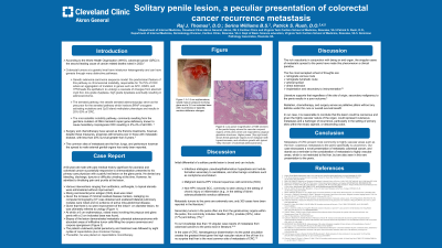
Introduction: Colorectal cancer (CRC) is the third leading cause of cancer-related deaths in the United States and 50% of CRC patients will develop metastases in the course of disease. Presented is a case of a 60-year-old male with a past medical history significant for psoriasis and colorectal cancer (Stage C T4bN1M0) treated by chemoradiation with complete response. A year later, the patient presented with painful red lesion on the penis clinically initially thought to be fungal balanitis, treated with various interventions ranging from antibiotics and antifungals without improvement. Biopsy of the lesion demonstrated metastatic colorectal adenocarcinoma filling the corpora. The patient went under partial penectomy and was advised to follow with chemoradiation. This case showcases an atypical presentation of metastatic colorectal cancer, and stands as a reminder to the possibility of metastasis to highly vascular areas, which are not restricted only the liver, but which also include the skin, particularly the scalp, and in this unusual presentation, the penis.
Case Description/Methods: A 60-year-old male with past medical history of psoriasis and rectal cancer successfully responsive to chemoradiation presented to with a painful red lesion on the glans penis. He denied any bleeding, discharge, dysuria or difficulty urinating. He admitted to throbbing pain at the area. He had no new sexual partners. Initially thought to be fungal balanitis. He was treated with various interventions ranging from antibiotics and antifungals without improvement. Given the progression of the penile lesion, he was referred to urology. On clinical exam, lesion showed advancement from initial presentation. A 6cm erythematous, raised mass in prepuce involving glans penis with a 2cm indurated base was found. Biopsy of the lesion demonstrated metastatic colorectal adenocarcinoma filling the corpora. The patient went under partial penectomy and was advised to follow with chemoradiation.
Discussion: Although there is a high propensity of colorectal cancer to metastasize to the liver, it is essential to keep in mind other vascular entities that may also be affected. This may include skin with a higher tendency to the scalp or as in this atypical presentation to the penis. However, given the past medical history of rectal carcinoma in this patient, it is reasonable to conclude that the lesion can be cancerous in nature and it should not deter one from making the diagnosis of metastatic rectal cancer in other highly vascular regions.

Disclosures:
Raj Jessica Thomas, DO1, Serina Williams, BS2, Patrick S. Rush, DO3. B0184 - Solitary Penile Lesion: A Peculiar Presentation of Colorectal Cancer Recurrence and Metastasis, ACG 2022 Annual Scientific Meeting Abstracts. Charlotte, NC: American College of Gastroenterology.
Raj Jessica Thomas, DO1, Serina Williams, BS2, Patrick S. Rush, DO3
1Cleveland Clinic Akron General, Akron, OH; 2Carilion Roanoke Memorial Hospital, Roanoke, VA; 3Virginia Tech Carilion School of Medicine, Roanoke, OH
Introduction: Colorectal cancer (CRC) is the third leading cause of cancer-related deaths in the United States and 50% of CRC patients will develop metastases in the course of disease. Presented is a case of a 60-year-old male with a past medical history significant for psoriasis and colorectal cancer (Stage C T4bN1M0) treated by chemoradiation with complete response. A year later, the patient presented with painful red lesion on the penis clinically initially thought to be fungal balanitis, treated with various interventions ranging from antibiotics and antifungals without improvement. Biopsy of the lesion demonstrated metastatic colorectal adenocarcinoma filling the corpora. The patient went under partial penectomy and was advised to follow with chemoradiation. This case showcases an atypical presentation of metastatic colorectal cancer, and stands as a reminder to the possibility of metastasis to highly vascular areas, which are not restricted only the liver, but which also include the skin, particularly the scalp, and in this unusual presentation, the penis.
Case Description/Methods: A 60-year-old male with past medical history of psoriasis and rectal cancer successfully responsive to chemoradiation presented to with a painful red lesion on the glans penis. He denied any bleeding, discharge, dysuria or difficulty urinating. He admitted to throbbing pain at the area. He had no new sexual partners. Initially thought to be fungal balanitis. He was treated with various interventions ranging from antibiotics and antifungals without improvement. Given the progression of the penile lesion, he was referred to urology. On clinical exam, lesion showed advancement from initial presentation. A 6cm erythematous, raised mass in prepuce involving glans penis with a 2cm indurated base was found. Biopsy of the lesion demonstrated metastatic colorectal adenocarcinoma filling the corpora. The patient went under partial penectomy and was advised to follow with chemoradiation.
Discussion: Although there is a high propensity of colorectal cancer to metastasize to the liver, it is essential to keep in mind other vascular entities that may also be affected. This may include skin with a higher tendency to the scalp or as in this atypical presentation to the penis. However, given the past medical history of rectal carcinoma in this patient, it is reasonable to conclude that the lesion can be cancerous in nature and it should not deter one from making the diagnosis of metastatic rectal cancer in other highly vascular regions.

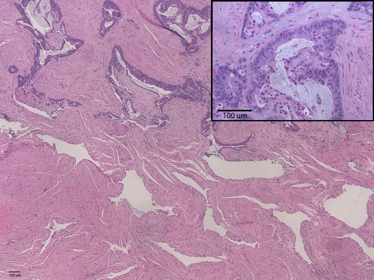
Figure: Low-power magnification of H&E sections of the penile sample shows the vascular corporal regions of the penis which are expanded by atypical glandular structures. Higher power (Top right inset) shows these glandular regions to be malignant with hyperchromasia, and cribriform growth with typical “dirty necrosis” of colorectal adenocarcinoma.
Disclosures:
Raj Jessica Thomas indicated no relevant financial relationships.
Serina Williams indicated no relevant financial relationships.
Patrick Rush indicated no relevant financial relationships.
Raj Jessica Thomas, DO1, Serina Williams, BS2, Patrick S. Rush, DO3. B0184 - Solitary Penile Lesion: A Peculiar Presentation of Colorectal Cancer Recurrence and Metastasis, ACG 2022 Annual Scientific Meeting Abstracts. Charlotte, NC: American College of Gastroenterology.

