Back


Poster Session A - Sunday Afternoon
Category: Colon
A0111 - A Rare Subtype of Colorectal Cancer Presenting as a Contained Perforation
Sunday, October 23, 2022
5:00 PM – 7:00 PM ET
Location: Crown Ballroom

Has Audio
- NP
Nishi K. Pandey, DO
CarePoint Health
Jersey City, NJ
Presenting Author(s)
Dhanush Hoskere, DO1, Melanie Burgos, MD2, Jennifer Hsieh, MD1, Andrew De La torre, MD3, Nishi K. Pandey, DO2
1CarePoint Health, Bayonne, NJ; 2CarePoint Health, Jersey City, NJ; 3CarePoint Health, Hoboken, NJ
Introduction: Colonic perforation comprises 3-10% of the initial presentation of Colorectal Cancer (CRC). Signet Ring Cell Carcinoma and Mucinous Adenocarcinoma are two distinct subtypes which occur with a very low incidence of 10-20% and 0.9-4%, respectively. Both are usually diagnosed at an advanced stage and have an overall worse prognosis than other types of CRC. Here we present a rare case of colonic mucinous adenocarcinoma with signet ring cell features manifesting as a contained perforated intra-abdominal mass necessitating surgery.
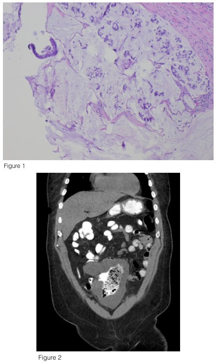
Case Description/Methods: A 60 year old male presented with fatigue, shortness of breath and anemia with hemoglobin of 4.8 g/dL. A computed tomography (CT) scan of the abdomen and pelvis was obtained which showed an intraperitoneal mass with a central necrotic component and air, suggesting communication to the small bowel. Due to the initial concern of a possible contained perforation, colonoscopy was deferred despite the patient never having one previously. Ultimately, the patient underwent a diagnostic laparoscopy which showed a large intra-abdominal mass with a mucinous surface involving multiple parts of the small bowel and sigmoid colon. This mass was removed along with a small bowel and sigmoid colon resection. Pathology was notable for moderately differentiated mucinous adenocarcinoma of the sigmoid colon with focal signet ring features with negative margins and no evidence of lymphovascular invasion. He was eventually discharged with instructions to follow up with Oncology for further treatment.
Discussion: Mucinous adenocarcinoma is characterized by the presence of abundant extracellular mucin which accounts for at least 50% of the tumor volume. Signet ring cells have copious amounts of mucin in the cytoplasm and nuclei located at the cell periphery. These lesions are usually located in the right colon and share similar characteristics on CT imaging including segments of concentric bowel wall thickening and ulcerations. Studies have shown that even a minor signet-ring cell component in CRC was associated with higher patient mortality. This becomes evident as these tumors diffusely infiltrate the submucosa with full thickness involvement of the bowel wall and have a high propensity for pelvic, peritoneal and ovarian metastasis. Treatment centers on surgical resection with adjuvant chemotherapy showing some benefit. Our case highlights an atypical presentation of a rare subtype of CRC for which gastroenterologists should maintain a high index of suspicion.

Disclosures:
Dhanush Hoskere, DO1, Melanie Burgos, MD2, Jennifer Hsieh, MD1, Andrew De La torre, MD3, Nishi K. Pandey, DO2. A0111 - A Rare Subtype of Colorectal Cancer Presenting as a Contained Perforation, ACG 2022 Annual Scientific Meeting Abstracts. Charlotte, NC: American College of Gastroenterology.
1CarePoint Health, Bayonne, NJ; 2CarePoint Health, Jersey City, NJ; 3CarePoint Health, Hoboken, NJ
Introduction: Colonic perforation comprises 3-10% of the initial presentation of Colorectal Cancer (CRC). Signet Ring Cell Carcinoma and Mucinous Adenocarcinoma are two distinct subtypes which occur with a very low incidence of 10-20% and 0.9-4%, respectively. Both are usually diagnosed at an advanced stage and have an overall worse prognosis than other types of CRC. Here we present a rare case of colonic mucinous adenocarcinoma with signet ring cell features manifesting as a contained perforated intra-abdominal mass necessitating surgery.
Case Description/Methods: A 60 year old male presented with fatigue, shortness of breath and anemia with hemoglobin of 4.8 g/dL. A computed tomography (CT) scan of the abdomen and pelvis was obtained which showed an intraperitoneal mass with a central necrotic component and air, suggesting communication to the small bowel. Due to the initial concern of a possible contained perforation, colonoscopy was deferred despite the patient never having one previously. Ultimately, the patient underwent a diagnostic laparoscopy which showed a large intra-abdominal mass with a mucinous surface involving multiple parts of the small bowel and sigmoid colon. This mass was removed along with a small bowel and sigmoid colon resection. Pathology was notable for moderately differentiated mucinous adenocarcinoma of the sigmoid colon with focal signet ring features with negative margins and no evidence of lymphovascular invasion. He was eventually discharged with instructions to follow up with Oncology for further treatment.
Discussion: Mucinous adenocarcinoma is characterized by the presence of abundant extracellular mucin which accounts for at least 50% of the tumor volume. Signet ring cells have copious amounts of mucin in the cytoplasm and nuclei located at the cell periphery. These lesions are usually located in the right colon and share similar characteristics on CT imaging including segments of concentric bowel wall thickening and ulcerations. Studies have shown that even a minor signet-ring cell component in CRC was associated with higher patient mortality. This becomes evident as these tumors diffusely infiltrate the submucosa with full thickness involvement of the bowel wall and have a high propensity for pelvic, peritoneal and ovarian metastasis. Treatment centers on surgical resection with adjuvant chemotherapy showing some benefit. Our case highlights an atypical presentation of a rare subtype of CRC for which gastroenterologists should maintain a high index of suspicion.

Figure: Figure 1. Signet Ring Cells in background of mucus (from sigmoid colon)
Figure 2. Computed Tomography (CT) scan showing an intra-abdominal mass with a contained perforation
Figure 2. Computed Tomography (CT) scan showing an intra-abdominal mass with a contained perforation
Disclosures:
Dhanush Hoskere indicated no relevant financial relationships.
Melanie Burgos indicated no relevant financial relationships.
Jennifer Hsieh indicated no relevant financial relationships.
Andrew De La torre indicated no relevant financial relationships.
Nishi Pandey indicated no relevant financial relationships.
Dhanush Hoskere, DO1, Melanie Burgos, MD2, Jennifer Hsieh, MD1, Andrew De La torre, MD3, Nishi K. Pandey, DO2. A0111 - A Rare Subtype of Colorectal Cancer Presenting as a Contained Perforation, ACG 2022 Annual Scientific Meeting Abstracts. Charlotte, NC: American College of Gastroenterology.
