Back
Poster Session A - Sunday Afternoon
A0696 - Malignant Transformation of a Gastric Duplication Cyst: A Rare Entity
Sunday, October 23, 2022
5:00 PM – 7:00 PM ET
Location: Crown Ballroom
.jpg)
Shuhaib S. Ali, DO
University of Texas Health San Antonio
San Antonio, TX
Presenting Author(s)
Award: Presidential Poster Award
Shuhaib Ali, DO1, Muhammad Haris, MD1, James Alvarez, MD2, Natasha McMillan, MD1, Laura Rosenkranz, MD1, Dhruv Mehta, MD3, Juan Echavarria, MS, MD1
1University of Texas Health San Antonio, San Antonio, TX; 2UT Health San Antonio, San Antonio, TX; 3University of Texas Health Science Center , San Antonio, TX
Introduction: Gastrointestinal tract duplication cysts (GTDCs) are rare congenital malformations that can occur anywhere along the alimentary tract. They occur most commonly in the ileum, esophagus, and colon. Gastric duplications cysts account for 4-9% of all intestinal duplication cysts. They are usually diagnosed at a young age due to their mass effect. Most cases in adults are incidentally discovered on endoscopy or radiological examination. Malignant transformation of duplications cysts in adults is extremely rare. We present a case of a gastric duplication cyst with malignant transformation.
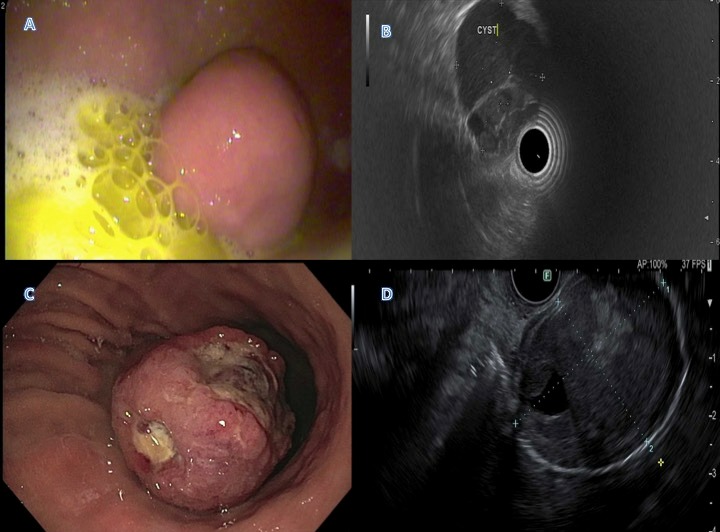
Case Description/Methods: A 69-year-old woman with a history of breast cancer status post resection and chemoradiation, left-sided ulcerative colitis well-controlled on mesalamine was incidentally found to have a gastric submucosal lesion (A) on esophagogastroduodenoscopy (EGD). Gastric biopsies were negative for Helicobacter Pylori. She subsequently underwent endoscopic ultrasound (EUS) with fine-needle aspiration (FNA) which revealed findings consistent with a gastric duplication cyst (B). FNA was negative for malignant cells. Eight years later she presented to the emergency room with melena and anemia. EGD revealed a large ulcerated subepithelial mass in the gastric body arising from the greater curvature; the area where earlier duplication cyst was found (C). EUS showed a hypoechoic round mass with isoechoic and anechoic components with well-defined borders (D). Gastric biopsies revealed poorly differentiated carcinoma and FNA was positive for malignant cells and signet ring cell carcinoma. Computed tomography of the abdomen and pelvis showed new hypodensities within the liver concerning for liver metastasis.
Discussion: This case demonstrates an unfortunate case of malignant transformation of a gastric duplication cyst. Malignant transformation of duplication cysts is extremely rare with only less than 15 cases reported in the literature. Based on prior studies and limited cases, no predictors for malignant change have been found including symptoms, size, location, tumor markers, or macroscopic findings. The mechanism for malignant transformation is poorly understood. If there is any suspicion of malignant transformation in the presence of a gastric duplication cyst, surgical resection is recommended.

Disclosures:
Shuhaib Ali, DO1, Muhammad Haris, MD1, James Alvarez, MD2, Natasha McMillan, MD1, Laura Rosenkranz, MD1, Dhruv Mehta, MD3, Juan Echavarria, MS, MD1. A0696 - Malignant Transformation of a Gastric Duplication Cyst: A Rare Entity, ACG 2022 Annual Scientific Meeting Abstracts. Charlotte, NC: American College of Gastroenterology.
Shuhaib Ali, DO1, Muhammad Haris, MD1, James Alvarez, MD2, Natasha McMillan, MD1, Laura Rosenkranz, MD1, Dhruv Mehta, MD3, Juan Echavarria, MS, MD1
1University of Texas Health San Antonio, San Antonio, TX; 2UT Health San Antonio, San Antonio, TX; 3University of Texas Health Science Center , San Antonio, TX
Introduction: Gastrointestinal tract duplication cysts (GTDCs) are rare congenital malformations that can occur anywhere along the alimentary tract. They occur most commonly in the ileum, esophagus, and colon. Gastric duplications cysts account for 4-9% of all intestinal duplication cysts. They are usually diagnosed at a young age due to their mass effect. Most cases in adults are incidentally discovered on endoscopy or radiological examination. Malignant transformation of duplications cysts in adults is extremely rare. We present a case of a gastric duplication cyst with malignant transformation.
Case Description/Methods: A 69-year-old woman with a history of breast cancer status post resection and chemoradiation, left-sided ulcerative colitis well-controlled on mesalamine was incidentally found to have a gastric submucosal lesion (A) on esophagogastroduodenoscopy (EGD). Gastric biopsies were negative for Helicobacter Pylori. She subsequently underwent endoscopic ultrasound (EUS) with fine-needle aspiration (FNA) which revealed findings consistent with a gastric duplication cyst (B). FNA was negative for malignant cells. Eight years later she presented to the emergency room with melena and anemia. EGD revealed a large ulcerated subepithelial mass in the gastric body arising from the greater curvature; the area where earlier duplication cyst was found (C). EUS showed a hypoechoic round mass with isoechoic and anechoic components with well-defined borders (D). Gastric biopsies revealed poorly differentiated carcinoma and FNA was positive for malignant cells and signet ring cell carcinoma. Computed tomography of the abdomen and pelvis showed new hypodensities within the liver concerning for liver metastasis.
Discussion: This case demonstrates an unfortunate case of malignant transformation of a gastric duplication cyst. Malignant transformation of duplication cysts is extremely rare with only less than 15 cases reported in the literature. Based on prior studies and limited cases, no predictors for malignant change have been found including symptoms, size, location, tumor markers, or macroscopic findings. The mechanism for malignant transformation is poorly understood. If there is any suspicion of malignant transformation in the presence of a gastric duplication cyst, surgical resection is recommended.

Figure: A) EGD demonstrating a gastric submucosal lesion in the body of the stomach
B) EUS illustrating 37 x 33 mm multiseptated anechoic lesion with internal debris
C) EGD findings showing a gastric mass occupying the entire lumen of distal gastric body with two overlying umbilicated ulcerations over the mass measuring 7 mm and 2 cm in size.
D) EUS demonstrating hypoechoic round mass of mixed features in the body of the stomach measuring 43 mm x 32 mm
Disclosures:
Shuhaib Ali indicated no relevant financial relationships.
Muhammad Haris indicated no relevant financial relationships.
James Alvarez indicated no relevant financial relationships.
Natasha McMillan indicated no relevant financial relationships.
Laura Rosenkranz indicated no relevant financial relationships.
Dhruv Mehta indicated no relevant financial relationships.
Juan Echavarria indicated no relevant financial relationships.
Shuhaib Ali, DO1, Muhammad Haris, MD1, James Alvarez, MD2, Natasha McMillan, MD1, Laura Rosenkranz, MD1, Dhruv Mehta, MD3, Juan Echavarria, MS, MD1. A0696 - Malignant Transformation of a Gastric Duplication Cyst: A Rare Entity, ACG 2022 Annual Scientific Meeting Abstracts. Charlotte, NC: American College of Gastroenterology.

