Back
Poster Session A - Sunday Afternoon
A0117 - Elderly Patient, Youthful Pathology: Solitary Colonic Juvenile Polyp Found in a 75-Year-Old Male
Sunday, October 23, 2022
5:00 PM – 7:00 PM ET
Location: Crown Ballroom

Praneeth Kudaravalli, MD
Augusta University Medical Center
Augusta, GA
Presenting Author(s)
Praneeth Kudaravalli, MD1, Christopher Moore, MS2, Intisar Ghleilib, MD3, Kenneth J. Vega, MD, FACG4, John Erikson L. Yap, MD4
1Augusta University Medical Center, Augusta, GA; 2Augusta University, Augusta, GA; 3Medical College of Georgia at Augusta University, Augusta, GA; 4Augusta University Medical College of Georiga, Augusta, GA
Introduction: Colonic juvenile polyps are generally found in the pediatric population. Classically, juvenile polyps (JP) are the most common polyp subtype seen in pediatric gastroenterology, accounting for approximately 80-90% of polyps in children. Generally, JP are diagnosed within the first decade of life and most common presentation is painless rectal bleeding. Other terminology used are juvenile hamartomatous polyp or retention polyps. There is no malignant potential for solitary JP and they tend to not recur.
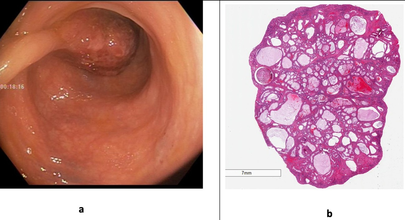
Case Description/Methods: We present a 75-year-old male with past medical history of hypertension, diabetes and hyperlipidemia referred to gastroenterology for surveillance colonoscopy due to a history of tubular adenomas. At the pre-procedure visit, the patient was well without GI complaints such as overt bleeding, change in bowel habits or unintentional weight loss. No family history of colon cancer or polyps was reported. Physical exam and vitals were normal at the time of procedure. Colonoscopy revealed a 40mm pedunculated sigmoid colon polyp which was excised using an endoloop at the stalk base followed by hot snare polypectomy (Fig. 1a). Three other sub-centimeter sessile polyps in the rectosigmoid area were removed by cold snare polypectomy. Histopathological examination of the large pedunculated polyp was consistent with a JP (Fig. 1b). Other polyps were tubular adenomas.
Discussion: Our current patient is the oldest reported case of isolated colonic JP. These polyps tend to have a common phenotypical appearance measuring around 10-30mm and roughly 90% of them are considered pedunculated. JP are ultimately defined by their distinctive histopathological features such as edematous lamina propria with inflammatory cells, cystically dilated glands that are bordered by cuboidal or columnar epithelium with reactive changes, and mucus filled glands. Fortunately for our patient, the presence of only a solitary colonic JP generally carries no known malignant potential. Juvenile polyposis syndrome (JPS) on the other hand is a hereditary condition characterized by the presence of hamartomatous polyps in the digestive tract and is known to have an increased risk of digestive tract malignancies. JPS is suspected when a patient has 5 or more juvenile polyps in the gastrointestinal tract or a juvenile polyp and a family history of juvenile polyps. A DNA test to check SMAD4 or BMPR1A gene is helpful.

Disclosures:
Praneeth Kudaravalli, MD1, Christopher Moore, MS2, Intisar Ghleilib, MD3, Kenneth J. Vega, MD, FACG4, John Erikson L. Yap, MD4. A0117 - Elderly Patient, Youthful Pathology: Solitary Colonic Juvenile Polyp Found in a 75-Year-Old Male, ACG 2022 Annual Scientific Meeting Abstracts. Charlotte, NC: American College of Gastroenterology.
1Augusta University Medical Center, Augusta, GA; 2Augusta University, Augusta, GA; 3Medical College of Georgia at Augusta University, Augusta, GA; 4Augusta University Medical College of Georiga, Augusta, GA
Introduction: Colonic juvenile polyps are generally found in the pediatric population. Classically, juvenile polyps (JP) are the most common polyp subtype seen in pediatric gastroenterology, accounting for approximately 80-90% of polyps in children. Generally, JP are diagnosed within the first decade of life and most common presentation is painless rectal bleeding. Other terminology used are juvenile hamartomatous polyp or retention polyps. There is no malignant potential for solitary JP and they tend to not recur.
Case Description/Methods: We present a 75-year-old male with past medical history of hypertension, diabetes and hyperlipidemia referred to gastroenterology for surveillance colonoscopy due to a history of tubular adenomas. At the pre-procedure visit, the patient was well without GI complaints such as overt bleeding, change in bowel habits or unintentional weight loss. No family history of colon cancer or polyps was reported. Physical exam and vitals were normal at the time of procedure. Colonoscopy revealed a 40mm pedunculated sigmoid colon polyp which was excised using an endoloop at the stalk base followed by hot snare polypectomy (Fig. 1a). Three other sub-centimeter sessile polyps in the rectosigmoid area were removed by cold snare polypectomy. Histopathological examination of the large pedunculated polyp was consistent with a JP (Fig. 1b). Other polyps were tubular adenomas.
Discussion: Our current patient is the oldest reported case of isolated colonic JP. These polyps tend to have a common phenotypical appearance measuring around 10-30mm and roughly 90% of them are considered pedunculated. JP are ultimately defined by their distinctive histopathological features such as edematous lamina propria with inflammatory cells, cystically dilated glands that are bordered by cuboidal or columnar epithelium with reactive changes, and mucus filled glands. Fortunately for our patient, the presence of only a solitary colonic JP generally carries no known malignant potential. Juvenile polyposis syndrome (JPS) on the other hand is a hereditary condition characterized by the presence of hamartomatous polyps in the digestive tract and is known to have an increased risk of digestive tract malignancies. JPS is suspected when a patient has 5 or more juvenile polyps in the gastrointestinal tract or a juvenile polyp and a family history of juvenile polyps. A DNA test to check SMAD4 or BMPR1A gene is helpful.

Figure: Figure 1. (a) A 40mm polyp (Paris 0-Ip) located in the in the sigmoid colon. (b) Hamartomatous polyp has ulcerated surface with benign dilated glands.
Disclosures:
Praneeth Kudaravalli indicated no relevant financial relationships.
Christopher Moore indicated no relevant financial relationships.
Intisar Ghleilib indicated no relevant financial relationships.
Kenneth Vega indicated no relevant financial relationships.
John Erikson Yap indicated no relevant financial relationships.
Praneeth Kudaravalli, MD1, Christopher Moore, MS2, Intisar Ghleilib, MD3, Kenneth J. Vega, MD, FACG4, John Erikson L. Yap, MD4. A0117 - Elderly Patient, Youthful Pathology: Solitary Colonic Juvenile Polyp Found in a 75-Year-Old Male, ACG 2022 Annual Scientific Meeting Abstracts. Charlotte, NC: American College of Gastroenterology.
