Back


Poster Session A - Sunday Afternoon
Category: GI Bleeding
A0305 - Pyogenic Granulomas: A Rare Cause of Small Bowel Bleeding
Sunday, October 23, 2022
5:00 PM – 7:00 PM ET
Location: Crown Ballroom

Has Audio
- WK
Whitney Kukol, MD
MetroHealth Medical Center/Case Western Reserve University
Cleveland, OH
Presenting Author(s)
Whitney Kukol, MD, Nisheet Waghray, MD
MetroHealth Medical Center/Case Western Reserve University, Cleveland, OH
Introduction: Bleeding from the small bowel is a relatively uncommon etiology of GI blood loss, accounting for only 5-10% of cases. Although advancements in video capsule endoscopy (VCE), enteroscopy, and angiography have assisted in identifying small bowel bleeds, their diagnosis and management remain challenging.
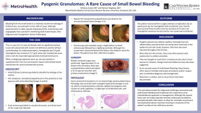
Case Description/Methods: This is a case of a 51-year-old female who presented with severe acute on chronic iron deficiency anemia requiring supplemental IV iron and multiple blood transfusions over the course of a year. She had no other significant medical issues. EGD and colonoscopy failed to identify the etiology of her anemia. VCE showed an ulcerated polypoid lesion in the proximal to mid jejunum with active bleeding. Push enteroscopy failed to visualize the lesion, and distal reach of the scope was tattooed. Repeat VCE showed the polypoid lesion just distal to the tattoo. Single balloon enteroscopy was performed; the scope was advanced beyond the tattoo, but failed to reach the lesion. The patient was referred to surgery and underwent robotic-assisted small bowel resection with primary anastomosis. Approximately 15 cm distal to the tattoo, there was evidence of a polypoid lesion prompting a 10 cm jejunal resection. Gross assessment revealed a 2.5 cm hemorrhagic pedunculated lesion with pathology consistent with a pyogenic granuloma. The patient recovered without complication, and, as of six weeks after surgery, her anemia had resolved.
Discussion: Pyogenic granulomas are inflammatory vascular lesions occurring most commonly in the epidermis and oral cavity; however, they have also been reported in the GI tract where they most often present with anemia. They are thought to result from a reactive process due to local trauma or irritation. As a rare cause of small bowel bleeding, they often require multiple endoscopic procedures and/or surgery prior to definitive diagnosis and management. Resection is curative, and no recurrences have been documented. This case demonstrates the diagnostic challenges associated with small bowel bleeding and emphasizes the importance of an interdisciplinary approach. Although this jejunal lesion was not endoscopically resected, endoscopic localization provided valuable information to allow for a minimally invasive and uncomplicated robotic resection resulting in resolution of the patient’s profound anemia.

Disclosures:
Whitney Kukol, MD, Nisheet Waghray, MD. A0305 - Pyogenic Granulomas: A Rare Cause of Small Bowel Bleeding, ACG 2022 Annual Scientific Meeting Abstracts. Charlotte, NC: American College of Gastroenterology.
MetroHealth Medical Center/Case Western Reserve University, Cleveland, OH
Introduction: Bleeding from the small bowel is a relatively uncommon etiology of GI blood loss, accounting for only 5-10% of cases. Although advancements in video capsule endoscopy (VCE), enteroscopy, and angiography have assisted in identifying small bowel bleeds, their diagnosis and management remain challenging.
Case Description/Methods: This is a case of a 51-year-old female who presented with severe acute on chronic iron deficiency anemia requiring supplemental IV iron and multiple blood transfusions over the course of a year. She had no other significant medical issues. EGD and colonoscopy failed to identify the etiology of her anemia. VCE showed an ulcerated polypoid lesion in the proximal to mid jejunum with active bleeding. Push enteroscopy failed to visualize the lesion, and distal reach of the scope was tattooed. Repeat VCE showed the polypoid lesion just distal to the tattoo. Single balloon enteroscopy was performed; the scope was advanced beyond the tattoo, but failed to reach the lesion. The patient was referred to surgery and underwent robotic-assisted small bowel resection with primary anastomosis. Approximately 15 cm distal to the tattoo, there was evidence of a polypoid lesion prompting a 10 cm jejunal resection. Gross assessment revealed a 2.5 cm hemorrhagic pedunculated lesion with pathology consistent with a pyogenic granuloma. The patient recovered without complication, and, as of six weeks after surgery, her anemia had resolved.
Discussion: Pyogenic granulomas are inflammatory vascular lesions occurring most commonly in the epidermis and oral cavity; however, they have also been reported in the GI tract where they most often present with anemia. They are thought to result from a reactive process due to local trauma or irritation. As a rare cause of small bowel bleeding, they often require multiple endoscopic procedures and/or surgery prior to definitive diagnosis and management. Resection is curative, and no recurrences have been documented. This case demonstrates the diagnostic challenges associated with small bowel bleeding and emphasizes the importance of an interdisciplinary approach. Although this jejunal lesion was not endoscopically resected, endoscopic localization provided valuable information to allow for a minimally invasive and uncomplicated robotic resection resulting in resolution of the patient’s profound anemia.

Figure: A. Ulcerated and bleeding polypoid lesion seen in jejunum on VCE. B. 10 cm jejunal resection with 2 roughly 1 cm polypoid lesions. C. Pathology specimen (described as a 2.5 x 1.2 x 1.2 cm pedunculated lesion) identified as a pyogenic granuloma.
Disclosures:
Whitney Kukol indicated no relevant financial relationships.
Nisheet Waghray indicated no relevant financial relationships.
Whitney Kukol, MD, Nisheet Waghray, MD. A0305 - Pyogenic Granulomas: A Rare Cause of Small Bowel Bleeding, ACG 2022 Annual Scientific Meeting Abstracts. Charlotte, NC: American College of Gastroenterology.
