Back


Poster Session C - Monday Afternoon
Category: Biliary/Pancreas
C0061 - An Extraordinary Case of Gallbladder Small Cell Carcinoma
Monday, October 24, 2022
3:00 PM – 5:00 PM ET
Location: Crown Ballroom

Has Audio

Ranbir Singh, MD
NYP Brooklyn Methodist Hospital
Brooklyn, NY
Presenting Author(s)
Ranbir Singh, MD1, Matthew Hanna, MD2
1NYP Brooklyn Methodist Hospital, Brooklyn, NY; 2Memorial Sloan Kettering Cancer Center, New York, NY
Introduction: We present a case of a patient diagnosed with Gallbladder Small Cell Carcinoma (SCC).
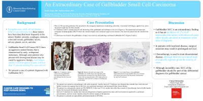
Case Description/Methods: This is a 58-year-old gentleman who presented to the emergency department complaining of pruritis. Associated with fatigue, appetite loss, and a 20-pound weight loss for three months. The physical exam was remarkable for scleral icterus and jaundice. Labs were remarkable for Total Bilirubin of 23.4 and Direct Bilirubin of 16.5. CT abdomen with IV contrast showed wall thickening of the gallbladder and infiltrates to his liver (Figure a). The patient had an Endoscopic retrograde cholangiography (ERCP) where the common hepatic duct contained single severe stenosis. One stent was placed into the common bile duct. A solid mass was found in the gallbladder, a biopsy was retrieved, and pathology confirmed Gallbladder SCC (Figures b and c). The patient was started on chemotherapy with carbo-etoposide. Another interval CT abdomen was obtained three months later which showed further invasion into segment four of the liver, new peritoneal metastasis, and increased biliary ductal dilation. The patient was subsequently started on FOLFIRINOX. Follow-up CT two months later after three rounds of chemotherapy showed stable hepatic lesions but increased intrahepatic ductal dilation. Interval CT abdomen two months later showed enlargement of hepatic segment four mass with involvement of segment five, peritoneal carcinomatosis, and new tumor implants along the anterior margin of the pancreas. The patient is currently on CAPTEM.
Discussion: Gallbladder SCC is an extraordinary finding as it has an incidence of .2% of all neuroendocrine tumors (1) with almost all cases either locally advanced or metastatic at the time of diagnosis. In Fuji et al’s case series review, it was found that the one-and two-year survival rates of the 53 cases were 28% and 0% respectively (2). In patients with localized disease, surgical resection may result in prolonged survival. Chemotherapy is used to treat disseminated disease, although there are no clinical trials of a specific regimen given the scarcity of cases. Although incredibly rare, SCC of the gallbladder should be part of the differential diagnosis for gallbladder cancer.
(1)Kanthan R, Senger JL, Ahmed S, et al. Gallbladder cancer in the 21st century. J.Oncol. 2015.
(2) Fujii H, Aotake T, Horiuchi T, et al. Small cell carcinoma of the gallbladder: a case report and review of 53 cases in the literature. Hepatogastroenterology. 2001.1588-93.

Disclosures:
Ranbir Singh, MD1, Matthew Hanna, MD2. C0061 - An Extraordinary Case of Gallbladder Small Cell Carcinoma, ACG 2022 Annual Scientific Meeting Abstracts. Charlotte, NC: American College of Gastroenterology.
1NYP Brooklyn Methodist Hospital, Brooklyn, NY; 2Memorial Sloan Kettering Cancer Center, New York, NY
Introduction: We present a case of a patient diagnosed with Gallbladder Small Cell Carcinoma (SCC).
Case Description/Methods: This is a 58-year-old gentleman who presented to the emergency department complaining of pruritis. Associated with fatigue, appetite loss, and a 20-pound weight loss for three months. The physical exam was remarkable for scleral icterus and jaundice. Labs were remarkable for Total Bilirubin of 23.4 and Direct Bilirubin of 16.5. CT abdomen with IV contrast showed wall thickening of the gallbladder and infiltrates to his liver (Figure a). The patient had an Endoscopic retrograde cholangiography (ERCP) where the common hepatic duct contained single severe stenosis. One stent was placed into the common bile duct. A solid mass was found in the gallbladder, a biopsy was retrieved, and pathology confirmed Gallbladder SCC (Figures b and c). The patient was started on chemotherapy with carbo-etoposide. Another interval CT abdomen was obtained three months later which showed further invasion into segment four of the liver, new peritoneal metastasis, and increased biliary ductal dilation. The patient was subsequently started on FOLFIRINOX. Follow-up CT two months later after three rounds of chemotherapy showed stable hepatic lesions but increased intrahepatic ductal dilation. Interval CT abdomen two months later showed enlargement of hepatic segment four mass with involvement of segment five, peritoneal carcinomatosis, and new tumor implants along the anterior margin of the pancreas. The patient is currently on CAPTEM.
Discussion: Gallbladder SCC is an extraordinary finding as it has an incidence of .2% of all neuroendocrine tumors (1) with almost all cases either locally advanced or metastatic at the time of diagnosis. In Fuji et al’s case series review, it was found that the one-and two-year survival rates of the 53 cases were 28% and 0% respectively (2). In patients with localized disease, surgical resection may result in prolonged survival. Chemotherapy is used to treat disseminated disease, although there are no clinical trials of a specific regimen given the scarcity of cases. Although incredibly rare, SCC of the gallbladder should be part of the differential diagnosis for gallbladder cancer.
(1)Kanthan R, Senger JL, Ahmed S, et al. Gallbladder cancer in the 21st century. J.Oncol. 2015.
(2) Fujii H, Aotake T, Horiuchi T, et al. Small cell carcinoma of the gallbladder: a case report and review of 53 cases in the literature. Hepatogastroenterology. 2001.1588-93.

Figure: Figure:
A: CT abdomen with IV contrast showing gallbladder wall thickening (black arrow) and infiltrates into the liver (red arrow).
B: Hematoxylin and eosin image of the gallbladder core needle biopsy showing high-grade neuroendocrine carcinoma involving fibrous stroma. Tumor cells are hyperchromatic with a high nucleus to cytoplasmic ratios and show evidence of molding and crush artifact.
C: Core needle biopsy of the gallbladder immunohistochemical stain for RB1 shows loss of tumor cell nuclear expression of the Rb protein, characteristic for small cell carcinoma
Disclosures:
Ranbir Singh indicated no relevant financial relationships.
Matthew Hanna indicated no relevant financial relationships.
Ranbir Singh, MD1, Matthew Hanna, MD2. C0061 - An Extraordinary Case of Gallbladder Small Cell Carcinoma, ACG 2022 Annual Scientific Meeting Abstracts. Charlotte, NC: American College of Gastroenterology.
