Back


Poster Session A - Sunday Afternoon
Category: Colon
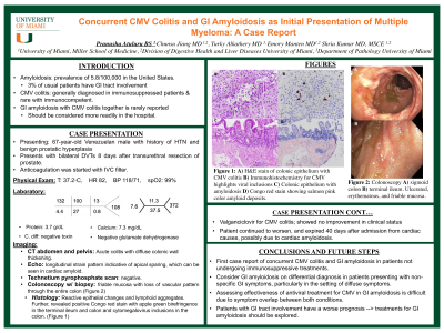
A0131 - Concurrent CMV Colitis and GI Amyloidosis as Initial Presentation of Multiple Myeloma: A Case Report
Sunday, October 23, 2022
5:00 PM – 7:00 PM ET
Location: Crown Ballroom

Has Audio
.jpeg.jpg)
Pranusha Atuluru, BS
University of Miami, Miller School of Medicine
Miami, FL
Presenting Author(s)
Pranusha Atuluru, BS1, Chunsu Jiang, MD2, Turky Alkathery, MD2, Emory Manten, MD2, Shria Kumar, MD2
1University of Miami, Miller School of Medicine, Miami, FL; 2Miller School of Medicine, University of Miami, Miami, FL
Introduction: Prior associations with Cytomegalovirus (CMV) colitis and multiple myeloma (MM) or amyloidosis are only described in immunocompromised patients undergoing treatments like transplantation, monoclonal antibodies, or chemotherapy. The concurrent presentation of gastrointestinal (GI) amyloidosis with CMV colitis is rarely reported and should be considered more readily in the hospital.
Case Description/Methods: 67-year-old male was admitted to the hospital with bilateral lower extremity deep vein thromboses after transurethral resection of the prostate 8 days prior. He was started on anticoagulation, but due to onset of hematuria, anticoagulation was stopped and an IVC filter was placed. The patient then developed bright red blood per rectum with diarrhea and GI was called. On exam, vitals were stable, and abdomen was non-distended and non-tender. Digital rectal exam showed brown stool. A colonoscopy displayed discontinuous areas of ulcerated, erythematous mucosa with loss of vascular pattern (1a). A histologic examination showed terminal ileal mucosa with lymphoid aggregates (1b), positive Congo red stain with apple green birefringence and cytomegalovirus inclusions in the colon (1c). Subsequent immunology testing demonstrated elevated free lambda light chains with a free kappa to lambda ratio of 0.019. A bone marrow biopsy confirmed lambda restriction and >20% CD138-positive plasma cells, consistent with MM. The patient was started on valganciclovir but showed no improvement of symptoms, which was attributed to his confounding amyloidosis. The patient’s clinical status continued to worsen, and he ultimately expired from cardiac causes.
Discussion: It is noteworthy that CMV colitis occurred concurrently with amyloidosis. Immune dysfunction and hypogammaglobinemia in amyloidosis lead to susceptibility to infection, but the inciting organisms usually are H. influenzae, S. pneumoniae, and E. coli. The use of immunosuppressing therapies such as transplantation or chemotherapy increases the likelihood of opportunistic infections. Besides these scenarios, CMV has rarely been reported in patients with amyloidosis or MM. We suggest GI amyloidosis and CMV should be considered and treated rapidly in patients presenting with non-specific GI symptoms. However, our case also highlights the difficulty in measuring effectiveness of antiviral treatment for CMV in GI amyloidosis due to overlap of symptoms between both conditions.

Disclosures:
Pranusha Atuluru, BS1, Chunsu Jiang, MD2, Turky Alkathery, MD2, Emory Manten, MD2, Shria Kumar, MD2. A0131 - Concurrent CMV Colitis and GI Amyloidosis as Initial Presentation of Multiple Myeloma: A Case Report, ACG 2022 Annual Scientific Meeting Abstracts. Charlotte, NC: American College of Gastroenterology.
1University of Miami, Miller School of Medicine, Miami, FL; 2Miller School of Medicine, University of Miami, Miami, FL
Introduction: Prior associations with Cytomegalovirus (CMV) colitis and multiple myeloma (MM) or amyloidosis are only described in immunocompromised patients undergoing treatments like transplantation, monoclonal antibodies, or chemotherapy. The concurrent presentation of gastrointestinal (GI) amyloidosis with CMV colitis is rarely reported and should be considered more readily in the hospital.
Case Description/Methods: 67-year-old male was admitted to the hospital with bilateral lower extremity deep vein thromboses after transurethral resection of the prostate 8 days prior. He was started on anticoagulation, but due to onset of hematuria, anticoagulation was stopped and an IVC filter was placed. The patient then developed bright red blood per rectum with diarrhea and GI was called. On exam, vitals were stable, and abdomen was non-distended and non-tender. Digital rectal exam showed brown stool. A colonoscopy displayed discontinuous areas of ulcerated, erythematous mucosa with loss of vascular pattern (1a). A histologic examination showed terminal ileal mucosa with lymphoid aggregates (1b), positive Congo red stain with apple green birefringence and cytomegalovirus inclusions in the colon (1c). Subsequent immunology testing demonstrated elevated free lambda light chains with a free kappa to lambda ratio of 0.019. A bone marrow biopsy confirmed lambda restriction and >20% CD138-positive plasma cells, consistent with MM. The patient was started on valganciclovir but showed no improvement of symptoms, which was attributed to his confounding amyloidosis. The patient’s clinical status continued to worsen, and he ultimately expired from cardiac causes.
Discussion: It is noteworthy that CMV colitis occurred concurrently with amyloidosis. Immune dysfunction and hypogammaglobinemia in amyloidosis lead to susceptibility to infection, but the inciting organisms usually are H. influenzae, S. pneumoniae, and E. coli. The use of immunosuppressing therapies such as transplantation or chemotherapy increases the likelihood of opportunistic infections. Besides these scenarios, CMV has rarely been reported in patients with amyloidosis or MM. We suggest GI amyloidosis and CMV should be considered and treated rapidly in patients presenting with non-specific GI symptoms. However, our case also highlights the difficulty in measuring effectiveness of antiviral treatment for CMV in GI amyloidosis due to overlap of symptoms between both conditions.

Figure: Figure 1: A) skiped areas of ulcerated, friable colon. B) immunohistochemistry CMV viral inclusions, C) Colonic epithelium with amyloidosis
Disclosures:
Pranusha Atuluru indicated no relevant financial relationships.
Chunsu Jiang indicated no relevant financial relationships.
Turky Alkathery indicated no relevant financial relationships.
Emory Manten indicated no relevant financial relationships.
Shria Kumar indicated no relevant financial relationships.
Pranusha Atuluru, BS1, Chunsu Jiang, MD2, Turky Alkathery, MD2, Emory Manten, MD2, Shria Kumar, MD2. A0131 - Concurrent CMV Colitis and GI Amyloidosis as Initial Presentation of Multiple Myeloma: A Case Report, ACG 2022 Annual Scientific Meeting Abstracts. Charlotte, NC: American College of Gastroenterology.
