Back


Poster Session B - Monday Morning
Category: Esophagus
B0248 - A Case of Severe Undifferentiated Ulcerative Esophagitis as an AIDS-Defining Illness in Acute HIV Infection
Monday, October 24, 2022
10:00 AM – 12:00 PM ET
Location: Crown Ballroom

Has Audio
- CW
Clemence White, DO
Walter Reed National Military Medical Center
Bethesda, MD
Presenting Author(s)
Clemence White, DO1, Christina Steinhauser, DO2
1Walter Reed National Military Medical Center, Bethesda, MD; 2Mike O’Callaghan Military Medical Center, Las Vegas, NV
Introduction: The esophagus is a common target organ in acute HIV infection (fungal or viral agents). Idiopathic esophageal ulcers are less common and we present a case of severe idiopathic ulcerative esophagitis in acute HIV infection.
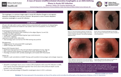
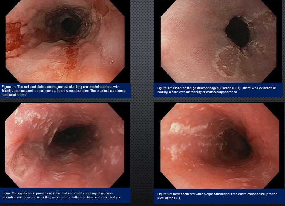
Case Description/Methods: 29-year old male with no prior medical history presented with symptoms of odynophagia, dysphagia and limited oral intake for two weeks. Due to his symptoms, he lost 25lbs at presentation. Esophagogastroduodenoscopy (EGD) revealed long cratered ulcerations with friability to the edges in the mid and distal esophagus (figure 1a). The mucosa between the ulcers appeared normal. Closer to the gastroesophageal junction (GEJ), there was evidence of healing ulcers without friability or cratered appearance (figure 1b). These lesions were biopsied (edges and base). The mucosa of the proximal esophagus, stomach and duodenum appeared normal. Due to endoscopic assessment concerning for viral infection, serology (HSV, CMV and HIV) were ordered. The patient was treated with empiric Valgancyclovir for presumed CMV esophagitis. The biopsy stained positive for undifferentiated hyphae type mold elements and viral stains were negative. His viral serology were normal but HIV RNA testing was positive. With infectious disease engagement, additional tests were done to rule out any opportunistic infection and results were negative. He was started on HAART therapy. Two weeks after initiating Valgancyclovir and HAART therapy, a repeat EGD showed significant improvement in the mid and distal esophageal mucosa ulceration with only one clean base cratered ulcer noted (Fig 2a). There were new and enlarged scattered white plaques throughout the entire esophagus up to the level of the GEJ (Fig. 2b). Several biopsies obtained from the ulcer and the white plaques were consistent with candida esophagitis, see figure 2b. He was treated with a course of fluconazole. Currently, patient is well controlled on HAART therapy with excellent immunologic and virologic response.
Discussion: Idiopathic esophageal ulcers occur in about 10% of patients with acute HIV infection or AIDS. Prior reports have described giant (greater than 5cm) ulcers with profound depth usually located in the mid esophagus. The exact pathophysiology is unknown. Gastroenterologists should maintain a low index suspicion for an immunocompromised state in all patients presenting with odynophagia or dysphagia. Upon diagnosis, compliance with HAART therapy is key for treatment of HIV related esophageal ulcers.

Disclosures:
Clemence White, DO1, Christina Steinhauser, DO2. B0248 - A Case of Severe Undifferentiated Ulcerative Esophagitis as an AIDS-Defining Illness in Acute HIV Infection, ACG 2022 Annual Scientific Meeting Abstracts. Charlotte, NC: American College of Gastroenterology.
1Walter Reed National Military Medical Center, Bethesda, MD; 2Mike O’Callaghan Military Medical Center, Las Vegas, NV
Introduction: The esophagus is a common target organ in acute HIV infection (fungal or viral agents). Idiopathic esophageal ulcers are less common and we present a case of severe idiopathic ulcerative esophagitis in acute HIV infection.
Case Description/Methods: 29-year old male with no prior medical history presented with symptoms of odynophagia, dysphagia and limited oral intake for two weeks. Due to his symptoms, he lost 25lbs at presentation. Esophagogastroduodenoscopy (EGD) revealed long cratered ulcerations with friability to the edges in the mid and distal esophagus (figure 1a). The mucosa between the ulcers appeared normal. Closer to the gastroesophageal junction (GEJ), there was evidence of healing ulcers without friability or cratered appearance (figure 1b). These lesions were biopsied (edges and base). The mucosa of the proximal esophagus, stomach and duodenum appeared normal. Due to endoscopic assessment concerning for viral infection, serology (HSV, CMV and HIV) were ordered. The patient was treated with empiric Valgancyclovir for presumed CMV esophagitis. The biopsy stained positive for undifferentiated hyphae type mold elements and viral stains were negative. His viral serology were normal but HIV RNA testing was positive. With infectious disease engagement, additional tests were done to rule out any opportunistic infection and results were negative. He was started on HAART therapy. Two weeks after initiating Valgancyclovir and HAART therapy, a repeat EGD showed significant improvement in the mid and distal esophageal mucosa ulceration with only one clean base cratered ulcer noted (Fig 2a). There were new and enlarged scattered white plaques throughout the entire esophagus up to the level of the GEJ (Fig. 2b). Several biopsies obtained from the ulcer and the white plaques were consistent with candida esophagitis, see figure 2b. He was treated with a course of fluconazole. Currently, patient is well controlled on HAART therapy with excellent immunologic and virologic response.
Discussion: Idiopathic esophageal ulcers occur in about 10% of patients with acute HIV infection or AIDS. Prior reports have described giant (greater than 5cm) ulcers with profound depth usually located in the mid esophagus. The exact pathophysiology is unknown. Gastroenterologists should maintain a low index suspicion for an immunocompromised state in all patients presenting with odynophagia or dysphagia. Upon diagnosis, compliance with HAART therapy is key for treatment of HIV related esophageal ulcers.

Disclosures:
Clemence White indicated no relevant financial relationships.
Christina Steinhauser indicated no relevant financial relationships.
Clemence White, DO1, Christina Steinhauser, DO2. B0248 - A Case of Severe Undifferentiated Ulcerative Esophagitis as an AIDS-Defining Illness in Acute HIV Infection, ACG 2022 Annual Scientific Meeting Abstracts. Charlotte, NC: American College of Gastroenterology.
