Back


Poster Session C - Monday Afternoon
Category: General Endoscopy
C0312 - Closure of Refractory Gastrocutaneous Fistula With Endoscopically Guided Percutaneous Suturing With the Use of SpyBite Biopsy Forceps
Monday, October 24, 2022
3:00 PM – 5:00 PM ET
Location: Crown Ballroom

Has Audio

Rogelio Garcia, MD
Baylor College of Medicine
Houston, TX
Presenting Author(s)
Rogelio Garcia, MD1, Juan D. Gomez Cifuentes, MD1, Tara Keihanian, MD1, Marvin Ryou, MD2, Wasif Abidi, MD, PhD1
1Baylor College of Medicine, Houston, TX; 2Brigham and Women's Hospital, Boston, MA
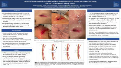
Introduction: Persistent gastrocutaneous fistula (GCF) is a rare but well-known complication of long-term Percutaneous Endoscopic Gastrostomy (PEG) tube use. To avoid invasive surgery, endoscopic closure has been used as an initial step for treatment but is not always successful. We report a case of successful GCF closure with a novel endoscopically guided percutaneous suturing technique using the SpyBite biopsy forceps.
Case Description/Methods: Our case is a 28-year-old male with a history of cystic fibrosis (CF) complicated by malnutrition, requiring PEG tube placement since childhood. After starting CF therapy with elexacaftor/tezacaftor/ivacaftor and achieving optimal nutritional status, his PEG tube was removed. Unfortunately, he developed a persistent GCF. Initial attempts at closure with over-the-scope-clip and endoscopic suturing failed. The decision was made to proceed with GCF closure by endoscopically guided percutaneous suturing using the SpyBite forceps.
Under endoscopic guidance, two 16G long angiocaths were advanced into the gastric lumen, one caudal and one cranial to the fistula tract in a sterile fashion. A 2-0 silk suture was advanced through one angiocath and externalized using SpyBite biopsy forceps through the other angiocath. The angiocaths were then removed over the suture and the loop was tied down using a surgical knot. This process was repeated two more times along the fistula tract, 5mm from each other. Internal closure of the GCF was then performed using endoscopic suturing. One interrupted and two running sutures were placed along the border and cinched to reinforce the site. There were no immediate adverse events or delayed skin inflammation. The patient had no further leakage from the GCF site at follow-up two weeks later.
Discussion: With the emergence of novel CF therapies, the dependence on feeding tubes has decreased. Unfortunately, these patients are at high risk of GCF formation after PEG tube removal. Given the difficulty in closing GCF, we advocate a multimodality approach, as described here, using transcutaneous and endoscopic suturing. In previously described endoscopically guided percutaneous suturing, the suture loop is externalized through the GCF tract or the mouth. Our technique differs in using SpyBite forceps to externalize the suture through a second catheter. This method is simple and provides a safe and effective alternative for the closure of refractory GCFs.

Disclosures:
Rogelio Garcia, MD1, Juan D. Gomez Cifuentes, MD1, Tara Keihanian, MD1, Marvin Ryou, MD2, Wasif Abidi, MD, PhD1. C0312 - Closure of Refractory Gastrocutaneous Fistula With Endoscopically Guided Percutaneous Suturing With the Use of SpyBite Biopsy Forceps, ACG 2022 Annual Scientific Meeting Abstracts. Charlotte, NC: American College of Gastroenterology.
1Baylor College of Medicine, Houston, TX; 2Brigham and Women's Hospital, Boston, MA
Introduction: Persistent gastrocutaneous fistula (GCF) is a rare but well-known complication of long-term Percutaneous Endoscopic Gastrostomy (PEG) tube use. To avoid invasive surgery, endoscopic closure has been used as an initial step for treatment but is not always successful. We report a case of successful GCF closure with a novel endoscopically guided percutaneous suturing technique using the SpyBite biopsy forceps.
Case Description/Methods: Our case is a 28-year-old male with a history of cystic fibrosis (CF) complicated by malnutrition, requiring PEG tube placement since childhood. After starting CF therapy with elexacaftor/tezacaftor/ivacaftor and achieving optimal nutritional status, his PEG tube was removed. Unfortunately, he developed a persistent GCF. Initial attempts at closure with over-the-scope-clip and endoscopic suturing failed. The decision was made to proceed with GCF closure by endoscopically guided percutaneous suturing using the SpyBite forceps.
Under endoscopic guidance, two 16G long angiocaths were advanced into the gastric lumen, one caudal and one cranial to the fistula tract in a sterile fashion. A 2-0 silk suture was advanced through one angiocath and externalized using SpyBite biopsy forceps through the other angiocath. The angiocaths were then removed over the suture and the loop was tied down using a surgical knot. This process was repeated two more times along the fistula tract, 5mm from each other. Internal closure of the GCF was then performed using endoscopic suturing. One interrupted and two running sutures were placed along the border and cinched to reinforce the site. There were no immediate adverse events or delayed skin inflammation. The patient had no further leakage from the GCF site at follow-up two weeks later.
Discussion: With the emergence of novel CF therapies, the dependence on feeding tubes has decreased. Unfortunately, these patients are at high risk of GCF formation after PEG tube removal. Given the difficulty in closing GCF, we advocate a multimodality approach, as described here, using transcutaneous and endoscopic suturing. In previously described endoscopically guided percutaneous suturing, the suture loop is externalized through the GCF tract or the mouth. Our technique differs in using SpyBite forceps to externalize the suture through a second catheter. This method is simple and provides a safe and effective alternative for the closure of refractory GCFs.

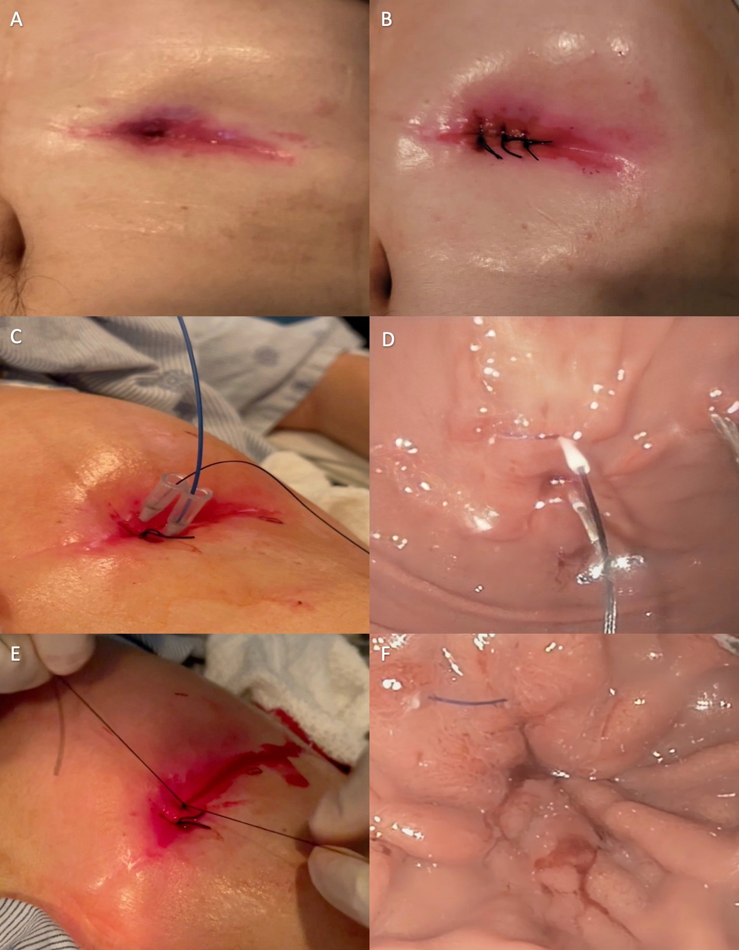
Figure: A-B: Gastrocutaneous fistula before (A) and after closure (B).
C-D: 2-0 silk suture inserted through the angiocath and SpyBite forceps inserted through the second angiocath. The suture was grasped with SpyBite forceps and pulled through the second angiocath to form a loop (C: external view, D: endoscopic view)
E-F: The suture was pulled externally, and a surgical knot was performed (E: external view, F: endoscopic view)
Disclosures:
Rogelio Garcia indicated no relevant financial relationships.
Juan D. Gomez Cifuentes indicated no relevant financial relationships.
Tara Keihanian indicated no relevant financial relationships.
Marvin Ryou: Boston Scientific – Consultant. Cook – Consultant, Grant/Research Support. EnteraSense – Consultant, Founder. Fujifilm – Consultant. GI windows – Consultant, Founder. Medtronic – Consultant. Olympus – Consultant.
Wasif Abidi: Ambu USA – Consultant. Apollo Endosurgery – Consultant. ConMEd – Consultant. GI dynamics – Grant/Research Support.
Rogelio Garcia, MD1, Juan D. Gomez Cifuentes, MD1, Tara Keihanian, MD1, Marvin Ryou, MD2, Wasif Abidi, MD, PhD1. C0312 - Closure of Refractory Gastrocutaneous Fistula With Endoscopically Guided Percutaneous Suturing With the Use of SpyBite Biopsy Forceps, ACG 2022 Annual Scientific Meeting Abstracts. Charlotte, NC: American College of Gastroenterology.

