Back


Poster Session D - Tuesday Morning
Category: GI Bleeding
D0327 - Omental Varices Causing Umbilical Bleeding: A Rare Presentation in Cirrhotic Patients With Portal Hypertension
Tuesday, October 25, 2022
10:00 AM – 12:00 PM ET
Location: Crown Ballroom

Has Audio

Genesis Perez Del Nogal, MD
TTUHSC
Odessa, Texas
Presenting Author(s)
Genesis Perez Del Nogal, MD1, Roman Karkee, MD1, Bibek Bakhati, MD2, Kalyan Chakrala, DO3
1TTUHSC, Odessa, TX; 2Texas Tech University Health Sciences Center, Odessa, TX; 3Medical Center Hospital, Odessa, TX
Introduction: Ectopic varices account for less than 5% of all varix-related bleeding episodes. Umbilical bleeding is a well-recognized but unusual complication of portal hypertension, and most of the time is caused by bleeding from umbilical varices (UV). On the other hand, rupture of omental varices (OV) leading to spontaneous umbilical bleeding is rare and has been reported once in the previous literature.
Case Description/Methods: A 54-year-old male with a history of liver cirrhosis, MELD score of 21, Child-Pugh Class B, who was found lying in a pool of blood in his bedroom, was transported to the hospital with profuse spontaneous bleeding from his umbilical hernia site. He gave a history of similar, but mild bleeding episodes occurring intermittently over the last week, with no trauma associated. Initially, the patient was hypotensive, tachycardic, and had a protuberant abdomen with a large ventral hernia, covered by a dressing. Removal of the dressing revealed active venous hemorrhage. Laboratories showed hemoglobin 6.9 g/dL, mean corpuscular volume 95.3 fL, platelets 88x103/mcL, INR 1.36, total bilirubin 0.7 mg/dL, ammonia level 160 µg/dL, and mild transaminitis. The patient’s hemorrhagic shock was treated with blood transfusions, IV fluids, and vasopressors. Abdomen CT scan showed anterior abdominal wall hernias containing herniated fat with fat stranding. The surgery service evaluated the patient and suspected OV bleeding. Intraoperatively, he was treated with periumbilical excision and ligation of the OV contained within the hernia. The omentum became adhered to the skin of the umbilicus, allowing venous communications to develop between the varices and cutaneous veins of the umbilicus. The section was dissected and a Seprafilm was placed to prevent future adherence, and the bleeding was controlled.
Discussion: The most reported cause of spontaneous umbilical bleeding is UV. In our case, the presence of bilateral anterior abdominal hernias containing herniated fat with some fat stranding on the abdominal CT was suspicious for OV, which was confirmed to be the source of bleeding intraoperatively. In such cases, OV as the source of umbilical bleeding should be ruled out. Previous reports have suggested a portosystemic abdominopelvic CT scan preoperatively to define the portal vascular anatomy, identify associated varices, and sites of bleeding before the procedure.

Disclosures:
Genesis Perez Del Nogal, MD1, Roman Karkee, MD1, Bibek Bakhati, MD2, Kalyan Chakrala, DO3. D0327 - Omental Varices Causing Umbilical Bleeding: A Rare Presentation in Cirrhotic Patients With Portal Hypertension, ACG 2022 Annual Scientific Meeting Abstracts. Charlotte, NC: American College of Gastroenterology.
1TTUHSC, Odessa, TX; 2Texas Tech University Health Sciences Center, Odessa, TX; 3Medical Center Hospital, Odessa, TX
Introduction: Ectopic varices account for less than 5% of all varix-related bleeding episodes. Umbilical bleeding is a well-recognized but unusual complication of portal hypertension, and most of the time is caused by bleeding from umbilical varices (UV). On the other hand, rupture of omental varices (OV) leading to spontaneous umbilical bleeding is rare and has been reported once in the previous literature.
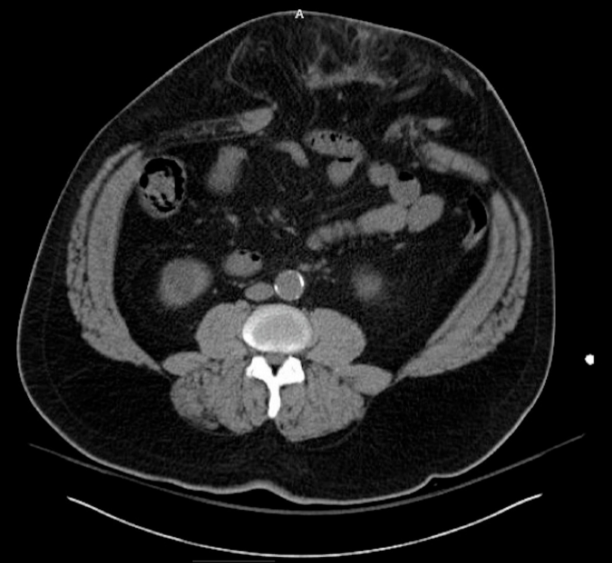
Case Description/Methods: A 54-year-old male with a history of liver cirrhosis, MELD score of 21, Child-Pugh Class B, who was found lying in a pool of blood in his bedroom, was transported to the hospital with profuse spontaneous bleeding from his umbilical hernia site. He gave a history of similar, but mild bleeding episodes occurring intermittently over the last week, with no trauma associated. Initially, the patient was hypotensive, tachycardic, and had a protuberant abdomen with a large ventral hernia, covered by a dressing. Removal of the dressing revealed active venous hemorrhage. Laboratories showed hemoglobin 6.9 g/dL, mean corpuscular volume 95.3 fL, platelets 88x103/mcL, INR 1.36, total bilirubin 0.7 mg/dL, ammonia level 160 µg/dL, and mild transaminitis. The patient’s hemorrhagic shock was treated with blood transfusions, IV fluids, and vasopressors. Abdomen CT scan showed anterior abdominal wall hernias containing herniated fat with fat stranding. The surgery service evaluated the patient and suspected OV bleeding. Intraoperatively, he was treated with periumbilical excision and ligation of the OV contained within the hernia. The omentum became adhered to the skin of the umbilicus, allowing venous communications to develop between the varices and cutaneous veins of the umbilicus. The section was dissected and a Seprafilm was placed to prevent future adherence, and the bleeding was controlled.
Discussion: The most reported cause of spontaneous umbilical bleeding is UV. In our case, the presence of bilateral anterior abdominal hernias containing herniated fat with some fat stranding on the abdominal CT was suspicious for OV, which was confirmed to be the source of bleeding intraoperatively. In such cases, OV as the source of umbilical bleeding should be ruled out. Previous reports have suggested a portosystemic abdominopelvic CT scan preoperatively to define the portal vascular anatomy, identify associated varices, and sites of bleeding before the procedure.

Figure: CT Abdomen and pelvis without contrast showing umbilical hernia containing omental fat.
Disclosures:
Genesis Perez Del Nogal indicated no relevant financial relationships.
Roman Karkee indicated no relevant financial relationships.
Bibek Bakhati indicated no relevant financial relationships.
Kalyan Chakrala indicated no relevant financial relationships.
Genesis Perez Del Nogal, MD1, Roman Karkee, MD1, Bibek Bakhati, MD2, Kalyan Chakrala, DO3. D0327 - Omental Varices Causing Umbilical Bleeding: A Rare Presentation in Cirrhotic Patients With Portal Hypertension, ACG 2022 Annual Scientific Meeting Abstracts. Charlotte, NC: American College of Gastroenterology.
