Back


Poster Session A - Sunday Afternoon
Category: Liver
A0584 - New Diagnosis of Systemic Amyloidosis in a Patient With Chronic Hepatitis B and Elevated Liver Stiffness Score on MR Elastography
Sunday, October 23, 2022
5:00 PM – 7:00 PM ET
Location: Crown Ballroom

Has Audio

Cristina I. Batarseh, MD
Lahey Hospital & Medical Center
Burlington, MA
Presenting Author(s)
Cristina I. Batarseh, MD, Atoussa Golder-Najafi, MD, Ali Khader, MD, Carmi S. Punzalan, MD
Lahey Hospital & Medical Center, Burlington, MA
Introduction: Amyloidosis is a disorder of abnormal deposition of protein into bodily tissue. This case describes a patient with chronic hepatitis B (HBV) who was found to have systemic amyloidosis after a magnetic resonance elastography (MRE) was consistent with advanced fibrosis. This case highlights the importance of confirming concerning MRE results with a liver biopsy.
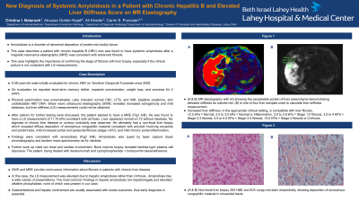
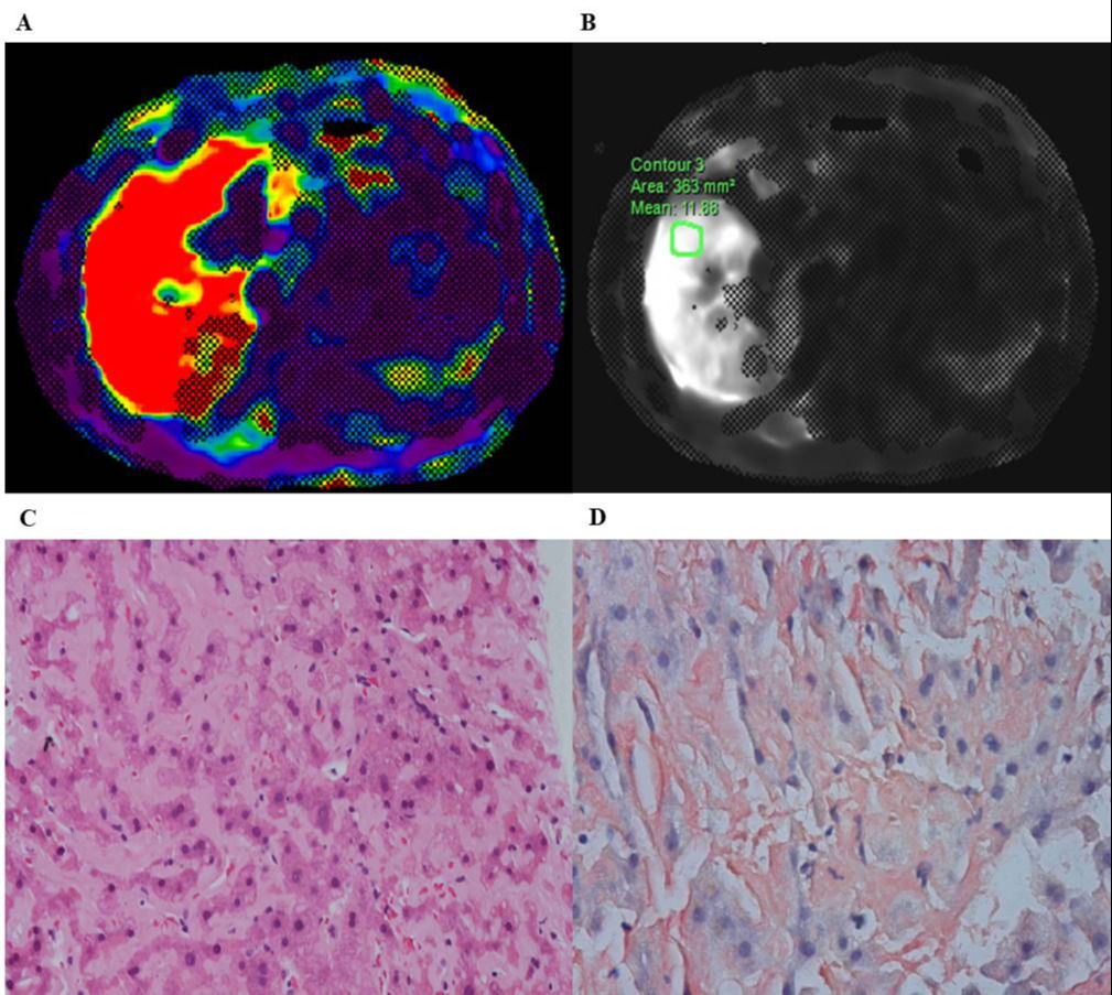
Case Description/Methods: A 58-year-old male initially evaluated for chronic HBV on Tenofovir Disoproxil Fumarate since 2009. He reported short-term memory defict, impaired concentration, weight loss, and anorexia for 2 years. Physical examination was unremarkable. Labs included normal CBC, LFTs, and INR, baseline creatinine, and undetectable HBV DNA. Shear wave ultrasound elastography (SWR) revealed increased echogenicity and mild steatosis, but liver stiffness (LS) measurements could not be obtained. After options for further testing were discussed, the patient elected to have a MRE (Fig1 A/B). He was found to have a LS measurement of 11.75 kPa consistent with cirrhosis. Liver appeared normal on T2 without steatosis. No stigmata of chronic liver disease or contour nodularity was observed. He ultimately had a non-focal liver biopsy which revealed diffuse deposition of amorphous congophilic material consistent with amyloid involving sinusoids and portal tracts, mild increased portal and periportal fibrosis (stage I-II/IV), and mild chronic portal inflammation. Findings were consistent with amyloidosis (Fig 1 C/D). Amyloidosis was typed by laser capture liquid chromatography and tandem mass spectrometry as AL–lambda. Further work up ruled out renal and cardiac involvement. Bone marrow biopsy revealed lambda-typic plasma cell dyscrasia. The patient is being treated with daratumumab and cyclophosphamide + bortezomib+dexamethasone.
Discussion: SWE and MRE provide noninvasive information about fibrosis in patients with chronic liver disease. This case highlights the importance of confirming the stage of fibrosis with liver biopsy, especially if the clinical picture is not consistent with LS measurements. In this case, the LS measurement was elevated due to hepatic amyloidosis rather than cirrhosis . Amyloidosis has a wide variety of presentations. The most common findings in hepatic amyloidosis are hepatomegaly and elevated alkaline phosphatase, none of which was present in our case. Gastrointestinal and hepatic involvement are usually associated with worse outcomes, thus early diagnosis is essential.

Disclosures:
Cristina I. Batarseh, MD, Atoussa Golder-Najafi, MD, Ali Khader, MD, Carmi S. Punzalan, MD. A0584 - New Diagnosis of Systemic Amyloidosis in a Patient With Chronic Hepatitis B and Elevated Liver Stiffness Score on MR Elastography, ACG 2022 Annual Scientific Meeting Abstracts. Charlotte, NC: American College of Gastroenterology.
Lahey Hospital & Medical Center, Burlington, MA
Introduction: Amyloidosis is a disorder of abnormal deposition of protein into bodily tissue. This case describes a patient with chronic hepatitis B (HBV) who was found to have systemic amyloidosis after a magnetic resonance elastography (MRE) was consistent with advanced fibrosis. This case highlights the importance of confirming concerning MRE results with a liver biopsy.
Case Description/Methods: A 58-year-old male initially evaluated for chronic HBV on Tenofovir Disoproxil Fumarate since 2009. He reported short-term memory defict, impaired concentration, weight loss, and anorexia for 2 years. Physical examination was unremarkable. Labs included normal CBC, LFTs, and INR, baseline creatinine, and undetectable HBV DNA. Shear wave ultrasound elastography (SWR) revealed increased echogenicity and mild steatosis, but liver stiffness (LS) measurements could not be obtained. After options for further testing were discussed, the patient elected to have a MRE (Fig1 A/B). He was found to have a LS measurement of 11.75 kPa consistent with cirrhosis. Liver appeared normal on T2 without steatosis. No stigmata of chronic liver disease or contour nodularity was observed. He ultimately had a non-focal liver biopsy which revealed diffuse deposition of amorphous congophilic material consistent with amyloid involving sinusoids and portal tracts, mild increased portal and periportal fibrosis (stage I-II/IV), and mild chronic portal inflammation. Findings were consistent with amyloidosis (Fig 1 C/D). Amyloidosis was typed by laser capture liquid chromatography and tandem mass spectrometry as AL–lambda. Further work up ruled out renal and cardiac involvement. Bone marrow biopsy revealed lambda-typic plasma cell dyscrasia. The patient is being treated with daratumumab and cyclophosphamide + bortezomib+dexamethasone.
Discussion: SWE and MRE provide noninvasive information about fibrosis in patients with chronic liver disease. This case highlights the importance of confirming the stage of fibrosis with liver biopsy, especially if the clinical picture is not consistent with LS measurements. In this case, the LS measurement was elevated due to hepatic amyloidosis rather than cirrhosis . Amyloidosis has a wide variety of presentations. The most common findings in hepatic amyloidosis are hepatomegaly and elevated alkaline phosphatase, none of which was present in our case. Gastrointestinal and hepatic involvement are usually associated with worse outcomes, thus early diagnosis is essential.

Figure: Figure 1. (A & B) MR elastography with (A) showing the sampleable portion of liver parenchyma demonstrating elevated stiffness as colored red. (B) is one of four liver samples used to calculate liver stiffness measurement. (C & D) Non-focal liver biopsy 20X H&E and 40X congo red stain respectively, showing deposition of amorphous congophilic material in sinusoidal tracts.
Disclosures:
Cristina Batarseh indicated no relevant financial relationships.
Atoussa Golder-Najafi indicated no relevant financial relationships.
Ali Khader indicated no relevant financial relationships.
Carmi Punzalan indicated no relevant financial relationships.
Cristina I. Batarseh, MD, Atoussa Golder-Najafi, MD, Ali Khader, MD, Carmi S. Punzalan, MD. A0584 - New Diagnosis of Systemic Amyloidosis in a Patient With Chronic Hepatitis B and Elevated Liver Stiffness Score on MR Elastography, ACG 2022 Annual Scientific Meeting Abstracts. Charlotte, NC: American College of Gastroenterology.
