Back

Poster Session A - Sunday Afternoon
Category: Small Intestine
A0680 - No Endoscopy, No Diagnosis: A Rare Presentation of Multiple Myeloma
Sunday, October 23, 2022
5:00 PM – 7:00 PM ET
Location: Crown Ballroom


Opeyemi Oluyemi, DO
Walter Reed National Military Medical Center
Bethesda, MD
Presenting Author(s)
Opeyemi Oluyemi, DO1, Andrew Mertz, MD1, Nhu-An Nguyen, DO2
1Walter Reed National Military Medical Center, Bethesda, MD; 2Joint Base Andrews, Andrews Air Force Base, MD
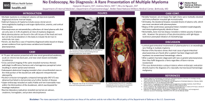
Introduction: Multiple myeloma (MM) is a malignant plasma cell dyscrasia typically found within the bone marrow, resulting in excess clonal serum immunoglobulins and ultimately end-organ dysfunction. Plasmacytomas are extramedullary collections of clonal plasma cell populations, which are only seen in 3.3% of patients at the time of MM diagnosis. Plasmacytomas are most commonly found in soft tissues of the head and neck and extremely rarely in the gastrointestinal system. Herein, we describe a case of MM diagnosed based solely on biopsy proven evidence of a singular vertebral plasmacytoma and a singular duodenal plasmacytoma.
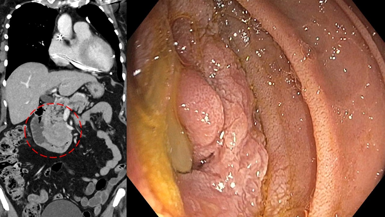
Case Description/Methods: A 73-year-old woman presented with generalized weakness, chronic back pain, increasing, and new onset incontinence. Spinal imaging revealed multiple thoracic pathologic vertebral insufficiency fractures and a homogenous paraspinal tumor resulting in severe spinal canal stenosis. Subsequent abdominal imaging demonstrated a large (6cm) circumferential mass involving the third portion of the duodenum with adjacent retroperitoneal adenopathy. An esophagogastroduodenoscopy was performed, revealing a non-traversable friable mass in the duodenum as the suspect location. Histologic evaluation revealed sheets of plasma cells extending to the duodenal mucosa with notably absent carcinoma. Routine laboratory assessment revealed normal calcium, creatinine, hemoglobin, and protein electrophoresis. However, serum kappa free light chains were elevated and immunofixation revealed IgA monoclonality. Biopsy of the synchronous spinal lesion revealed morphologically and immunophenotypically similar tissue. Interestingly, bone marrow biopsy revealed a paucity of plasma cells. The patient underwent surgical spinal cord decompression and promptly started chemotherapy.
Discussion: Gastrointestinal involvement is a rare finding in MM and is almost universally seen after the patient has been diagnosed via conventional criteria. Typically, this diagnosis is made when plasma cells comprise >10% of bone marrow specimen along with typical clinical features. This patient solely demonstrated lytic axial bone lesions with a bone marrow biopsy revealing too few plasma cells to complete immunohistochemical analysis. However, in the presence of >1 plasmacytomas, bone marrow involvement is not essential to make the diagnosis. This case demonstrates a unique instance when endoscopic evaluation was instrumental in the diagnosis of a classically marrow-based hematologic malignancy.

Disclosures:
Opeyemi Oluyemi, DO1, Andrew Mertz, MD1, Nhu-An Nguyen, DO2. A0680 - No Endoscopy, No Diagnosis: A Rare Presentation of Multiple Myeloma, ACG 2022 Annual Scientific Meeting Abstracts. Charlotte, NC: American College of Gastroenterology.
1Walter Reed National Military Medical Center, Bethesda, MD; 2Joint Base Andrews, Andrews Air Force Base, MD
Introduction: Multiple myeloma (MM) is a malignant plasma cell dyscrasia typically found within the bone marrow, resulting in excess clonal serum immunoglobulins and ultimately end-organ dysfunction. Plasmacytomas are extramedullary collections of clonal plasma cell populations, which are only seen in 3.3% of patients at the time of MM diagnosis. Plasmacytomas are most commonly found in soft tissues of the head and neck and extremely rarely in the gastrointestinal system. Herein, we describe a case of MM diagnosed based solely on biopsy proven evidence of a singular vertebral plasmacytoma and a singular duodenal plasmacytoma.
Case Description/Methods: A 73-year-old woman presented with generalized weakness, chronic back pain, increasing, and new onset incontinence. Spinal imaging revealed multiple thoracic pathologic vertebral insufficiency fractures and a homogenous paraspinal tumor resulting in severe spinal canal stenosis. Subsequent abdominal imaging demonstrated a large (6cm) circumferential mass involving the third portion of the duodenum with adjacent retroperitoneal adenopathy. An esophagogastroduodenoscopy was performed, revealing a non-traversable friable mass in the duodenum as the suspect location. Histologic evaluation revealed sheets of plasma cells extending to the duodenal mucosa with notably absent carcinoma. Routine laboratory assessment revealed normal calcium, creatinine, hemoglobin, and protein electrophoresis. However, serum kappa free light chains were elevated and immunofixation revealed IgA monoclonality. Biopsy of the synchronous spinal lesion revealed morphologically and immunophenotypically similar tissue. Interestingly, bone marrow biopsy revealed a paucity of plasma cells. The patient underwent surgical spinal cord decompression and promptly started chemotherapy.
Discussion: Gastrointestinal involvement is a rare finding in MM and is almost universally seen after the patient has been diagnosed via conventional criteria. Typically, this diagnosis is made when plasma cells comprise >10% of bone marrow specimen along with typical clinical features. This patient solely demonstrated lytic axial bone lesions with a bone marrow biopsy revealing too few plasma cells to complete immunohistochemical analysis. However, in the presence of >1 plasmacytomas, bone marrow involvement is not essential to make the diagnosis. This case demonstrates a unique instance when endoscopic evaluation was instrumental in the diagnosis of a classically marrow-based hematologic malignancy.

Figure: Large ill-defined mass adjacent to the third portion of the duodenum; seen on esophagogastroduodenoscopy as a circumferential, non-traversable and friable lesion, consistent with plasma cell neoplasm on tissue examination.
Disclosures:
Opeyemi Oluyemi indicated no relevant financial relationships.
Andrew Mertz indicated no relevant financial relationships.
Nhu-An Nguyen indicated no relevant financial relationships.
Opeyemi Oluyemi, DO1, Andrew Mertz, MD1, Nhu-An Nguyen, DO2. A0680 - No Endoscopy, No Diagnosis: A Rare Presentation of Multiple Myeloma, ACG 2022 Annual Scientific Meeting Abstracts. Charlotte, NC: American College of Gastroenterology.
