Back


Poster Session D - Tuesday Morning
Category: Liver
D0544 - Hepatocellular Carcinoma in a Non-Cirrhotic Patient Treated With Left Hepatic Trisegmentectomy: A Case Report
Tuesday, October 25, 2022
10:00 AM – 12:00 PM ET
Location: Crown Ballroom

Has Audio

James S. Love, MD
University of Illinois at Chicago
Chicago, IL
Presenting Author(s)
James S. Love, MD1, Sarang Thaker, MD, MS1, Alexander Pan, MD1, Josi Herren, DO1, Alberto Fratti, MD1, Grace Guzman, MD2, Mario Spaggiari, MD1, Sean Koppe, MD1
1University of Illinois at Chicago, Chicago, IL; 2University of Illinois Medical Center, Chicago, IL
Introduction: Hepatocellular carcinoma (HCC) is the 5th-most common cancer and the 3rd-most common cause of cancer-related mortality. Chronic liver disease is the most important risk factor for HCC and 80% of cases are in patients with cirrhosis. HCC has an insidious presentation in patients without cirrhosis and is often found incidentally.
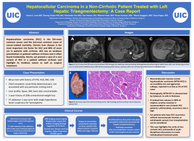
Case Description/Methods: A 68-year-old male presented with acute right upper quadrant pain. History was notable only for daily 12-beer consumption for the preceding 6 months. Laboratory tests revealed normal liver profile and international normalized ratio. Ultrasound demonstrated hepatomegaly and a heterogenous mass. Computed tomography (CT) with contrast of the abdomen demonstrated a large mass involving segments 2, 3, 4, 5, and 8 measuring 19.8 cm in largest dimension. Magnetic resonance imaging was concerning for fibrolamellar variant HCC without imaging findings suggestive of cirrhosis (fig. 1). CT scan from 1 year prior demonstrated non-cirrhotic liver morphology and hemangioma in the left lobe. Chronic liver disease workup was unremarkable. Alpha-fetoprotein was elevated to 150,000 ng/mL. Staging scans were negative for metastatic disease. Biopsy of normal liver parenchyma excluded underlying cirrhosis with mild mixed vesicular steatosis (20%) and stage 2 fibrosis. Volumetry confirmed adequate residual liver volume. After multi-disciplinary discussions, the patient underwent a left hepatic trisegmentecomy. Pathology demonstrated macrotrabecular-massive variant of HCC with lymphovascular invasion (Fig. 2).
Discussion: Macrotrabecular-massive variant is characterized by thick trabeculae and has been reported in as few as 5% of HCC cases. It is associated with frequent lymphovascular invasion and elevated AFP levels. If residual liver volume is ≥40% of original, curative resection is recommended in non-cirrhotic HCC patients. Unfortunately, post-resection recurrence rate is high, and risk factors include microvascular invasion, large tumor size, AFP >400 ng/mL, and cirrhosis. For patients who have HCC recurrence without macrovascular invasion or extrahepatic spread, liver transplant may be considered. The Barcelona Clinic Liver Cancer staging system and the Liver Imaging Reporting and Data System (LI-RADS) have been widely validated in patients with cirrhosis; however, neither system has been studied in non-cirrhotic HCC. This case highlights the rarity of non-cirrhotic HCC and benefit of multi-disciplinary discussions to create individualized treatment plans.

Disclosures:
James S. Love, MD1, Sarang Thaker, MD, MS1, Alexander Pan, MD1, Josi Herren, DO1, Alberto Fratti, MD1, Grace Guzman, MD2, Mario Spaggiari, MD1, Sean Koppe, MD1. D0544 - Hepatocellular Carcinoma in a Non-Cirrhotic Patient Treated With Left Hepatic Trisegmentectomy: A Case Report, ACG 2022 Annual Scientific Meeting Abstracts. Charlotte, NC: American College of Gastroenterology.
1University of Illinois at Chicago, Chicago, IL; 2University of Illinois Medical Center, Chicago, IL
Introduction: Hepatocellular carcinoma (HCC) is the 5th-most common cancer and the 3rd-most common cause of cancer-related mortality. Chronic liver disease is the most important risk factor for HCC and 80% of cases are in patients with cirrhosis. HCC has an insidious presentation in patients without cirrhosis and is often found incidentally.
Case Description/Methods: A 68-year-old male presented with acute right upper quadrant pain. History was notable only for daily 12-beer consumption for the preceding 6 months. Laboratory tests revealed normal liver profile and international normalized ratio. Ultrasound demonstrated hepatomegaly and a heterogenous mass. Computed tomography (CT) with contrast of the abdomen demonstrated a large mass involving segments 2, 3, 4, 5, and 8 measuring 19.8 cm in largest dimension. Magnetic resonance imaging was concerning for fibrolamellar variant HCC without imaging findings suggestive of cirrhosis (fig. 1). CT scan from 1 year prior demonstrated non-cirrhotic liver morphology and hemangioma in the left lobe. Chronic liver disease workup was unremarkable. Alpha-fetoprotein was elevated to 150,000 ng/mL. Staging scans were negative for metastatic disease. Biopsy of normal liver parenchyma excluded underlying cirrhosis with mild mixed vesicular steatosis (20%) and stage 2 fibrosis. Volumetry confirmed adequate residual liver volume. After multi-disciplinary discussions, the patient underwent a left hepatic trisegmentecomy. Pathology demonstrated macrotrabecular-massive variant of HCC with lymphovascular invasion (Fig. 2).
Discussion: Macrotrabecular-massive variant is characterized by thick trabeculae and has been reported in as few as 5% of HCC cases. It is associated with frequent lymphovascular invasion and elevated AFP levels. If residual liver volume is ≥40% of original, curative resection is recommended in non-cirrhotic HCC patients. Unfortunately, post-resection recurrence rate is high, and risk factors include microvascular invasion, large tumor size, AFP >400 ng/mL, and cirrhosis. For patients who have HCC recurrence without macrovascular invasion or extrahepatic spread, liver transplant may be considered. The Barcelona Clinic Liver Cancer staging system and the Liver Imaging Reporting and Data System (LI-RADS) have been widely validated in patients with cirrhosis; however, neither system has been studied in non-cirrhotic HCC. This case highlights the rarity of non-cirrhotic HCC and benefit of multi-disciplinary discussions to create individualized treatment plans.

Figure: Figure 1: (A) Coronal and (B) axial arterial phase MRI through the abdomen demonstrating heterogenously enhancing bi-lobed mass with non-enhancing central scar. (C) Axial portal venous phase MRI demonstrating portal venous washout with non-enhancing central scars in a bilobed mass.
Figure 2: (A) Gross specimen demonstrating tumor. (B) Histology demonstrating macrotrabecular variant HCC.
Figure 2: (A) Gross specimen demonstrating tumor. (B) Histology demonstrating macrotrabecular variant HCC.
Disclosures:
James Love indicated no relevant financial relationships.
Sarang Thaker indicated no relevant financial relationships.
Alexander Pan indicated no relevant financial relationships.
Josi Herren indicated no relevant financial relationships.
Alberto Fratti indicated no relevant financial relationships.
Grace Guzman indicated no relevant financial relationships.
Mario Spaggiari indicated no relevant financial relationships.
Sean Koppe indicated no relevant financial relationships.
James S. Love, MD1, Sarang Thaker, MD, MS1, Alexander Pan, MD1, Josi Herren, DO1, Alberto Fratti, MD1, Grace Guzman, MD2, Mario Spaggiari, MD1, Sean Koppe, MD1. D0544 - Hepatocellular Carcinoma in a Non-Cirrhotic Patient Treated With Left Hepatic Trisegmentectomy: A Case Report, ACG 2022 Annual Scientific Meeting Abstracts. Charlotte, NC: American College of Gastroenterology.
