Back


Poster Session D - Tuesday Morning
Category: Stomach
D0712 - Collagenous Gastritis: A Rare Cause of Severe Iron Deficiency Anemia in a Basic Military Trainee
Tuesday, October 25, 2022
10:00 AM – 12:00 PM ET
Location: Crown Ballroom

Has Audio
- KS
Kendra T. Stilwell, DO
San Antonio Uniformed Services Health Education Consortium
San Antonio, TX
Presenting Author(s)
Kendra T. Stilwell, DO1, Douglas B. Walton, MD2, James M. Francis, DO2, Charles B. Miller, MD2, Geoffrey A. Bader, MD2
1San Antonio Uniformed Services Health Education Consortium, San Antonio, TX; 2SAUSHEC, San Antonio, TX
Introduction: Collagenous gastritis is a rare inflammatory disease characterized by deposition of a thick subepithelial band of
collagen and associated inflammatory infiltrate. This disorder has a reported prevalence of 13 per 100,000
esophagogastroduodenoscopies (EGDs) with a female predominance. Two distinct clinical phenotypes have been
described: adult-onset characterized by diffuse gastrointestinal involvement and diarrhea, and pediatric-onset
manifesting with abdominal pain and anemia. We present an interesting case of pediatric-phenotype collagenous
gastritis to highlight this poorly-recognized disorder.
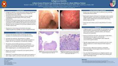
Case Description/Methods: A 19-year-old male basic military trainee was referred for evaluation of incidentally discovered iron deficiency
anemia. He denied any symptoms, overt blood loss, or NSAIDs. Physical exam was unremarkable. Labs revealed
hemoglobin of 9.3g/dL, iron saturation 7% and ferritin 15ng/mL. The patient underwent EGD and colonoscopy for
further evaluation. EGD revealed a diffusely nodular appearance of the gastric mucosa with areas with suspected
atrophy under narrow-band imaging (Figures A, B). Colonoscopy was normal. Biopsies returned with a non-specific
chronic gastritis without H. pylori. Abdominal computed tomography and additional work up for autoimmune
gastritis, fecal H. pylori antigen, syphilis, and heavy metals toxins were all normal. Decision was made to repeat
EGD for additional tissue sampling, including cold snared samples. Repeat gastric biopsies with expert GI
pathologist review revealed collagenous gastritis, characterized by subepithelial deposition of a thick (greater than 10µm)
collagen band and associated inflammatory infiltrate (Figures C, D). Patient was lost to follow up due to
disqualification for military service.
Discussion: Collagenous gastritis is an exceedingly rare heterogeneous disease process of poorly understood causes and
pathogenesis, with autoimmune conditions, medication effects, and infections theorized to be responsible. Numerous
treatments have been reported with variable effect, including antisecretory agents, corticosteroids,
immunomodulators, and hypoallergenic diets, along with micronutrient supplementation. Due to its rarity, a high
level of suspicion is required by gastroenterologists and pathologists based on clinical and endoscopic findings. Our
case helps to bring awareness to collagenous gastritis, and emphasizes the potential value for repeat tissue sampling
with expert pathologist review.

Disclosures:
Kendra T. Stilwell, DO1, Douglas B. Walton, MD2, James M. Francis, DO2, Charles B. Miller, MD2, Geoffrey A. Bader, MD2. D0712 - Collagenous Gastritis: A Rare Cause of Severe Iron Deficiency Anemia in a Basic Military Trainee, ACG 2022 Annual Scientific Meeting Abstracts. Charlotte, NC: American College of Gastroenterology.
1San Antonio Uniformed Services Health Education Consortium, San Antonio, TX; 2SAUSHEC, San Antonio, TX
Introduction: Collagenous gastritis is a rare inflammatory disease characterized by deposition of a thick subepithelial band of
collagen and associated inflammatory infiltrate. This disorder has a reported prevalence of 13 per 100,000
esophagogastroduodenoscopies (EGDs) with a female predominance. Two distinct clinical phenotypes have been
described: adult-onset characterized by diffuse gastrointestinal involvement and diarrhea, and pediatric-onset
manifesting with abdominal pain and anemia. We present an interesting case of pediatric-phenotype collagenous
gastritis to highlight this poorly-recognized disorder.
Case Description/Methods: A 19-year-old male basic military trainee was referred for evaluation of incidentally discovered iron deficiency
anemia. He denied any symptoms, overt blood loss, or NSAIDs. Physical exam was unremarkable. Labs revealed
hemoglobin of 9.3g/dL, iron saturation 7% and ferritin 15ng/mL. The patient underwent EGD and colonoscopy for
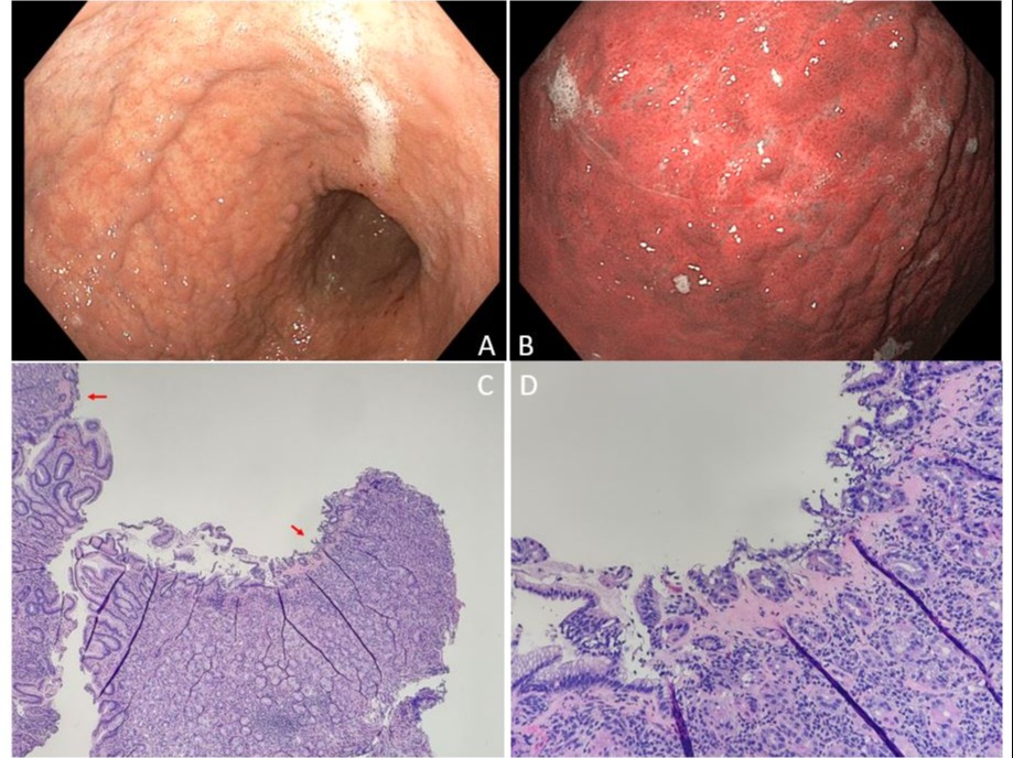
further evaluation. EGD revealed a diffusely nodular appearance of the gastric mucosa with areas with suspected
atrophy under narrow-band imaging (Figures A, B). Colonoscopy was normal. Biopsies returned with a non-specific
chronic gastritis without H. pylori. Abdominal computed tomography and additional work up for autoimmune
gastritis, fecal H. pylori antigen, syphilis, and heavy metals toxins were all normal. Decision was made to repeat
EGD for additional tissue sampling, including cold snared samples. Repeat gastric biopsies with expert GI
pathologist review revealed collagenous gastritis, characterized by subepithelial deposition of a thick (greater than 10µm)
collagen band and associated inflammatory infiltrate (Figures C, D). Patient was lost to follow up due to
disqualification for military service.
Discussion: Collagenous gastritis is an exceedingly rare heterogeneous disease process of poorly understood causes and
pathogenesis, with autoimmune conditions, medication effects, and infections theorized to be responsible. Numerous
treatments have been reported with variable effect, including antisecretory agents, corticosteroids,
immunomodulators, and hypoallergenic diets, along with micronutrient supplementation. Due to its rarity, a high
level of suspicion is required by gastroenterologists and pathologists based on clinical and endoscopic findings. Our
case helps to bring awareness to collagenous gastritis, and emphasizes the potential value for repeat tissue sampling
with expert pathologist review.

Figure: A: Diffusely nodular appearance of the gastric mucosa with areas. B: Narrow-band imaging reveals areas of suspected atrophy. C: Multiple fragments of gastric mucosa with a prominent lymphoplasmacytic infiltrate within the lamina propria and occasional foci of subepithelial collagen deposition (red arrows). [Hematoxylin & Eosin, 50X magnification]. D: On higher magnification a focus of subepithelial collagen deposition may be seen associated with epithelial injury indicated by loss of foveolar mucin and detachment of the surface epithelium. [Hematoxylin & Eosin, 200X magnification]
Disclosures:
Kendra Stilwell indicated no relevant financial relationships.
Douglas Walton indicated no relevant financial relationships.
James Francis indicated no relevant financial relationships.
Charles Miller indicated no relevant financial relationships.
Geoffrey Bader indicated no relevant financial relationships.
Kendra T. Stilwell, DO1, Douglas B. Walton, MD2, James M. Francis, DO2, Charles B. Miller, MD2, Geoffrey A. Bader, MD2. D0712 - Collagenous Gastritis: A Rare Cause of Severe Iron Deficiency Anemia in a Basic Military Trainee, ACG 2022 Annual Scientific Meeting Abstracts. Charlotte, NC: American College of Gastroenterology.
