Back


Poster Session D - Tuesday Morning
Category: GI Bleeding
D0339 - Pseudoaneurysm: A Very Real GI Bleed
Tuesday, October 25, 2022
10:00 AM – 12:00 PM ET
Location: Crown Ballroom

Has Audio

Hajira Malik, MD
University of South Alabama
Mobile, Alabama
Presenting Author(s)
Hajira Zafar Malik, MD, E. Baylee Edwards, , Abrahim Hanjar, MD, Rajab Idriss, MD
University of South Alabama, Mobile, AL
Introduction: There are different causes and a broad spectrum of clinical presentations of upper gastrointestinal bleeding (UGIB). There are some rare causes of UGIB that can prove fatal if not identified and treated early. We present the case of a patient with known history of alcohol abuse disorder and recurrent acute pancreatitis complicated by splenic artery pseudoaneurysm causing GI bleed.
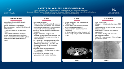
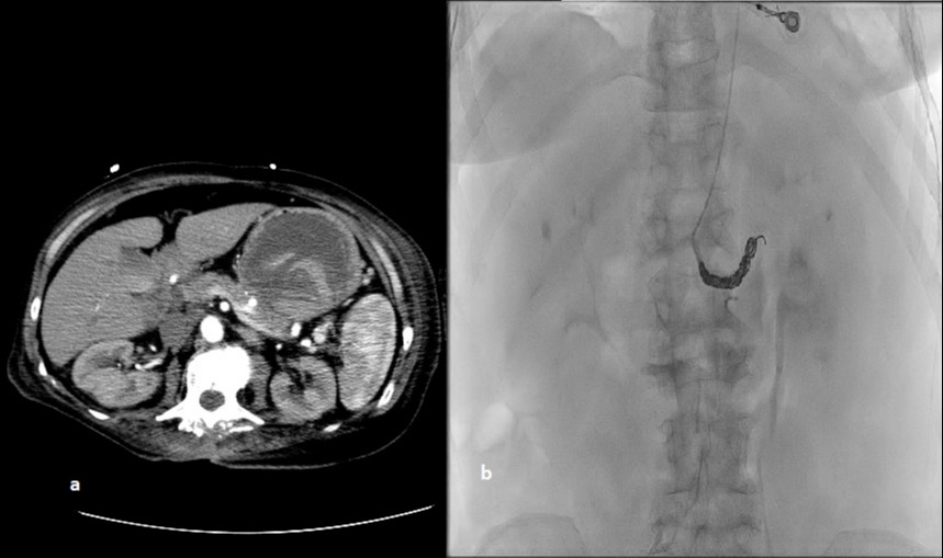
Case Description/Methods: 60 years old female with past medical history of hepatitis C, recurrent acute alcoholic pancreatitis and liver cirrhosis was hospitalized for administration of intravenous antibiotics to treat infected back wound. On day 12 of hospitalization, she developed hematemesis with hemodynamic instability. She underwent esophagogastroduodenoscopy (EGD) showing a large >5 cm protuberant soft mass-like lesion with superficial ulceration and no active bleed in the gastric fundus. Appearance was concerning for a vascular lesion and CT abdomen was done. CT showed interval enlargement of a presumed previously demonstrated pancreatic pseudocyst measuring 8.6x9.1x11.1cm. Density of the pseudocyst contents on CT ranged between 52-56 Hounsfield units consistent with recent bleeding (Image 1a). Patient developed hematochezia after which CT angiogram with GI protocol was done which did not show active extravasation. Based on imaging, it was initially assumed that patient had hemorrhaged into the presumed pseudocyst that had eroded into the wall of fundus, as seen in the EGD. Patient underwent arterial angiogram with Interventional Radiology (IR). Splenic arteriogram found a splenic artery pseudoaneurysm, instead of a pseudocyst, with new active contrast extravasation supplied by splenic artery. The pseudoaneurysm was most likely secondary to acute recurrent pancreatitis. She underwent Gel foam coil embolization of splenic artery (Image 1b) with resolution of UGIB.
Discussion: Bleeding from splenic artery pseudoaneurysm is very rare with less than 200 cases of splenic artery pseudoaneurysm reported in literature. It may present as an upper or lower GI bleed, or as in our case, both. Suspicion is reasonable in a patient with history of pancreatitis. Bleeding from splenic artery pseudoaneurysm is essential to identify early to reduce morbidity and mortality. This case brings attention to an infrequent yet life-threatening source of GI bleed which may be overlooked as it is rarely encountered and emphasizes the role of IR in management of hemorrhagic pseudoaneurysms.

Disclosures:
Hajira Zafar Malik, MD, E. Baylee Edwards, , Abrahim Hanjar, MD, Rajab Idriss, MD. D0339 - Pseudoaneurysm: A Very Real GI Bleed, ACG 2022 Annual Scientific Meeting Abstracts. Charlotte, NC: American College of Gastroenterology.
University of South Alabama, Mobile, AL
Introduction: There are different causes and a broad spectrum of clinical presentations of upper gastrointestinal bleeding (UGIB). There are some rare causes of UGIB that can prove fatal if not identified and treated early. We present the case of a patient with known history of alcohol abuse disorder and recurrent acute pancreatitis complicated by splenic artery pseudoaneurysm causing GI bleed.
Case Description/Methods: 60 years old female with past medical history of hepatitis C, recurrent acute alcoholic pancreatitis and liver cirrhosis was hospitalized for administration of intravenous antibiotics to treat infected back wound. On day 12 of hospitalization, she developed hematemesis with hemodynamic instability. She underwent esophagogastroduodenoscopy (EGD) showing a large >5 cm protuberant soft mass-like lesion with superficial ulceration and no active bleed in the gastric fundus. Appearance was concerning for a vascular lesion and CT abdomen was done. CT showed interval enlargement of a presumed previously demonstrated pancreatic pseudocyst measuring 8.6x9.1x11.1cm. Density of the pseudocyst contents on CT ranged between 52-56 Hounsfield units consistent with recent bleeding (Image 1a). Patient developed hematochezia after which CT angiogram with GI protocol was done which did not show active extravasation. Based on imaging, it was initially assumed that patient had hemorrhaged into the presumed pseudocyst that had eroded into the wall of fundus, as seen in the EGD. Patient underwent arterial angiogram with Interventional Radiology (IR). Splenic arteriogram found a splenic artery pseudoaneurysm, instead of a pseudocyst, with new active contrast extravasation supplied by splenic artery. The pseudoaneurysm was most likely secondary to acute recurrent pancreatitis. She underwent Gel foam coil embolization of splenic artery (Image 1b) with resolution of UGIB.
Discussion: Bleeding from splenic artery pseudoaneurysm is very rare with less than 200 cases of splenic artery pseudoaneurysm reported in literature. It may present as an upper or lower GI bleed, or as in our case, both. Suspicion is reasonable in a patient with history of pancreatitis. Bleeding from splenic artery pseudoaneurysm is essential to identify early to reduce morbidity and mortality. This case brings attention to an infrequent yet life-threatening source of GI bleed which may be overlooked as it is rarely encountered and emphasizes the role of IR in management of hemorrhagic pseudoaneurysms.

Figure: a: CT abdomen demonstrating the presumed pseudocyst. b: Gel foam coil embolization of splenic artery
Disclosures:
Hajira Zafar Malik indicated no relevant financial relationships.
E. Baylee Edwards indicated no relevant financial relationships.
Abrahim Hanjar indicated no relevant financial relationships.
Rajab Idriss indicated no relevant financial relationships.
Hajira Zafar Malik, MD, E. Baylee Edwards, , Abrahim Hanjar, MD, Rajab Idriss, MD. D0339 - Pseudoaneurysm: A Very Real GI Bleed, ACG 2022 Annual Scientific Meeting Abstracts. Charlotte, NC: American College of Gastroenterology.
