Back


Poster Session E - Tuesday Afternoon
Category: Biliary/Pancreas
E0068 - Acinar Cell Cystadenoma With Pancreatic Intraepithelial Neoplasia: Is It Always Benign?
Tuesday, October 25, 2022
3:00 PM – 5:00 PM ET
Location: Crown Ballroom

Has Audio

Somtochukwu Onwuzo, MD
Cleveland Clinic Foundation
Cleveland, OH
Presenting Author(s)
Somtochukwu Onwuzo, MD, Hassan M. Shaheen, MD, Ashraf Almomani, MD, Eduard Krishtopaytis, MD, Prabhat Kumar, MD, Antoine Boustany, MD, MPH, Ala Abdel Jalil, MD
Cleveland Clinic Foundation, Cleveland, OH
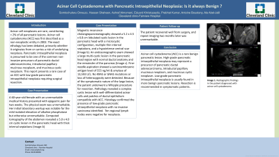
Introduction: Acinar cell neoplasms are rare, constituting < 2% of all pancreatic lesions. Acinar cell cystadenoma (ACC) was first described as a non-neoplastic entity in 2002. The exact etiology has been debated, primarily whether it originates from or carries a risk of underlying neoplasia. Pancreatic intraepithelial neoplasia (PanIN) is presumed to be one of the common noninvasive precursors of pancreatic ductal adenocarcinoma, intraductal papillary mucinous neoplasm, and mucinous cystic neoplasm. This report presents a rare case of an ACC with low-grade PanIN requiring surgical resection.
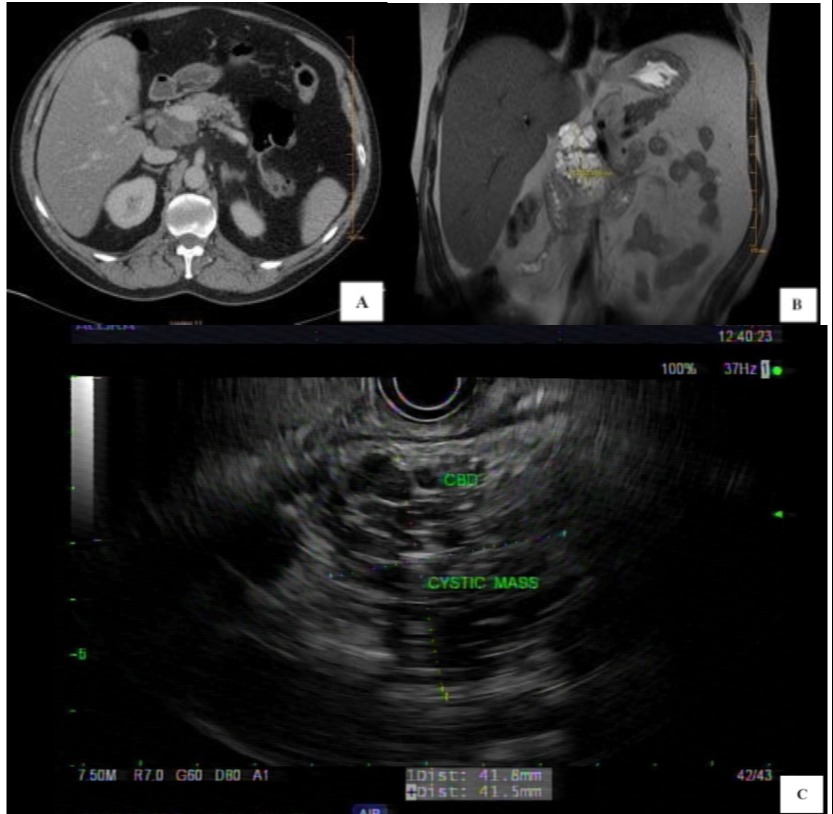
Case Description/Methods: A 60-year-old female with an unremarkable medical history presented with epigastric pain for two weeks. The physical exam was unremarkable. Her initial laboratory workup was notable for the mild isolated elevation of alkaline phosphatase but otherwise unremarkable. Computed tomography of the abdomen revealed a 5.0 x 4.0 cm cystic lesion in the pancreatic head with thick internal septations (Image a). Magnetic resonance cholangiopancreatography showed a 5.2 x 4.5 x 6.8 cm lobulated cystic lesion in the pancreatic head with a microcystic configuration, multiple thin internal septations, and a hypointense central scar (Image b). An endosonographic exam showed a large multi-cystic lesion in the pancreatic head region with normal ductal anatomy and the remainder of the pancreas (Image c). Fine-needle aspiration showed a carcinoembryonic antigen level of 555 ng/ml & amylase of 13,593 U/L. No KRAS or GNAS mutations or loss of heterozygosity were detected. Because of the symptomatic nature of the large lesion, the patient underwent a Whipple procedure for resection. Pathology revealed a complex cystic lesion with well-differentiated acinar cells and patches of ductal epithelium compatible with ACC. Histology confirmed the presence of low-grade pancreatic intraepithelial neoplasia (PanIN) with no invasive carcinoma identified. Ten regional lymph nodes were negative for neoplasia. The patient recovered well from surgery, and repeat imaging two months later was unremarkable.
Discussion: Acinar cell cystadenoma (ACC) is a rare benign pancreatic lesion. High-grade pancreatic intraepithelial neoplasia (PanIN) may represent a precursor of pancreatic ductal adenocarcinoma, intraductal papillary mucinous neoplasm, and mucinous cystic neoplasm. Low-grade PanIN is usually found in more benign pancreatic lesions. Resection is recommended in symptomatic patients.

Disclosures:
Somtochukwu Onwuzo, MD, Hassan M. Shaheen, MD, Ashraf Almomani, MD, Eduard Krishtopaytis, MD, Prabhat Kumar, MD, Antoine Boustany, MD, MPH, Ala Abdel Jalil, MD. E0068 - Acinar Cell Cystadenoma With Pancreatic Intraepithelial Neoplasia: Is It Always Benign?, ACG 2022 Annual Scientific Meeting Abstracts. Charlotte, NC: American College of Gastroenterology.
Cleveland Clinic Foundation, Cleveland, OH
Introduction: Acinar cell neoplasms are rare, constituting < 2% of all pancreatic lesions. Acinar cell cystadenoma (ACC) was first described as a non-neoplastic entity in 2002. The exact etiology has been debated, primarily whether it originates from or carries a risk of underlying neoplasia. Pancreatic intraepithelial neoplasia (PanIN) is presumed to be one of the common noninvasive precursors of pancreatic ductal adenocarcinoma, intraductal papillary mucinous neoplasm, and mucinous cystic neoplasm. This report presents a rare case of an ACC with low-grade PanIN requiring surgical resection.
Case Description/Methods: A 60-year-old female with an unremarkable medical history presented with epigastric pain for two weeks. The physical exam was unremarkable. Her initial laboratory workup was notable for the mild isolated elevation of alkaline phosphatase but otherwise unremarkable. Computed tomography of the abdomen revealed a 5.0 x 4.0 cm cystic lesion in the pancreatic head with thick internal septations (Image a). Magnetic resonance cholangiopancreatography showed a 5.2 x 4.5 x 6.8 cm lobulated cystic lesion in the pancreatic head with a microcystic configuration, multiple thin internal septations, and a hypointense central scar (Image b). An endosonographic exam showed a large multi-cystic lesion in the pancreatic head region with normal ductal anatomy and the remainder of the pancreas (Image c). Fine-needle aspiration showed a carcinoembryonic antigen level of 555 ng/ml & amylase of 13,593 U/L. No KRAS or GNAS mutations or loss of heterozygosity were detected. Because of the symptomatic nature of the large lesion, the patient underwent a Whipple procedure for resection. Pathology revealed a complex cystic lesion with well-differentiated acinar cells and patches of ductal epithelium compatible with ACC. Histology confirmed the presence of low-grade pancreatic intraepithelial neoplasia (PanIN) with no invasive carcinoma identified. Ten regional lymph nodes were negative for neoplasia. The patient recovered well from surgery, and repeat imaging two months later was unremarkable.
Discussion: Acinar cell cystadenoma (ACC) is a rare benign pancreatic lesion. High-grade pancreatic intraepithelial neoplasia (PanIN) may represent a precursor of pancreatic ductal adenocarcinoma, intraductal papillary mucinous neoplasm, and mucinous cystic neoplasm. Low-grade PanIN is usually found in more benign pancreatic lesions. Resection is recommended in symptomatic patients.

Figure: Radiographic findings in the patient diagnosed with acinar cell cystadenoma
Disclosures:
Somtochukwu Onwuzo indicated no relevant financial relationships.
Hassan Shaheen indicated no relevant financial relationships.
Ashraf Almomani indicated no relevant financial relationships.
Eduard Krishtopaytis indicated no relevant financial relationships.
Prabhat Kumar indicated no relevant financial relationships.
Antoine Boustany indicated no relevant financial relationships.
Ala Abdel Jalil indicated no relevant financial relationships.
Somtochukwu Onwuzo, MD, Hassan M. Shaheen, MD, Ashraf Almomani, MD, Eduard Krishtopaytis, MD, Prabhat Kumar, MD, Antoine Boustany, MD, MPH, Ala Abdel Jalil, MD. E0068 - Acinar Cell Cystadenoma With Pancreatic Intraepithelial Neoplasia: Is It Always Benign?, ACG 2022 Annual Scientific Meeting Abstracts. Charlotte, NC: American College of Gastroenterology.
