Back


Poster Session E - Tuesday Afternoon
Category: Biliary/Pancreas
E0075 - A Case of Biliary Intraepithelial Neoplasia of the Extrahepatic Bile Duct
Tuesday, October 25, 2022
3:00 PM – 5:00 PM ET
Location: Crown Ballroom

Has Audio

Sonya Bhaskar, MD
University of South Florida
Tampa, Florida
Presenting Author(s)
Sonya Bhaskar, MD, Ali Zakaria, MD, Pushpak Taunk, MD
University of South Florida, Tampa, FL
Introduction: Biliary intraepithelial neoplasia (BilIN) is a relatively new diagnosis that refers to flat or micropapillary precursor lesions of the bile duct which can develop into adenocarcinomas. Herein, we present a case of an extrahepatic biliary intraepithelial neoplasia that was found during endoscopic retrograde cholangiopancreatography (ERCP).
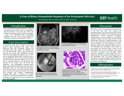
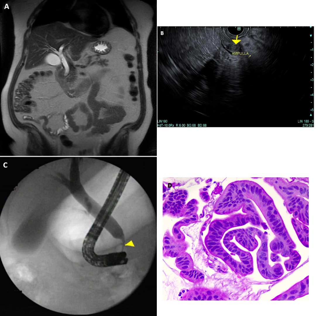
Case Description/Methods: 54-year-old male with a history of hypertension and tobacco abuse presented with sharp, right upper quadrant abdominal pain with intermittent nausea and vomiting of one month duration. On admission, he was hemodynamically stable, with normal labs including liver function tests. MRI/MRCP revealed intra- and extra-hepatic duct dilation up to 12 mm with no intraductal filling defect or stricture and a normal gallbladder. [Fig. 1A].The patient underwent an EUS which revealed common bile duct dilation up to 12 mm with a hypoechoic lesion in the distal bile duct, just proximal to the ampulla [Fig. 1B]. Given the EUS findings, an ERCP was performed. Cholangiogram revealed diffuse dilation of the biliary tree with no obvious stricture or filling defect. [Fig. 1C]. The biliary tree was swept, and nothing was found. Due to the suspicious lesion seen on the EUS we performed distal common bile duct brushing and biopsies. Histopathology revealed biliary intraepithelial neoplasia, grade 2. [Fig.1D].His pain was controlled and was discharged with plans for pancreaticoduodenectomy (Whipple’s) procedure.
Discussion: BilIN refers to flat or micropapillary precursor lesions of the bile duct which can develop into adenocarcinomas. They are commonly found in liver samples of chronic biliary and liver diseases such as hepatolithiasis, primary sclerosing cholangitis, choledochal cyst, chronic hepatitis C, and alcoholic cirrhosis. However, they are rarely found in clinical practice since they are not easily accessible, nor do they usually cause bile duct obstruction. The natural course of BillN is not well understood and in 2017, diagnostic criteria were created for lesions found in the intrahepatic duct based on their cellular and structural atypia. Lesions in the extrahepatic duct do occur, however, very rarely. This case is unusual in that the BilIN caused symptoms suggestive of biliary obstruction with a dilated bile duct and because the BillN was found in the extrahepatic duct. The case highlights the importance of considering BilIN in biliary obstruction and perform biopsies to identify the lesions prior to development of malignancy.

Disclosures:
Sonya Bhaskar, MD, Ali Zakaria, MD, Pushpak Taunk, MD. E0075 - A Case of Biliary Intraepithelial Neoplasia of the Extrahepatic Bile Duct, ACG 2022 Annual Scientific Meeting Abstracts. Charlotte, NC: American College of Gastroenterology.
University of South Florida, Tampa, FL
Introduction: Biliary intraepithelial neoplasia (BilIN) is a relatively new diagnosis that refers to flat or micropapillary precursor lesions of the bile duct which can develop into adenocarcinomas. Herein, we present a case of an extrahepatic biliary intraepithelial neoplasia that was found during endoscopic retrograde cholangiopancreatography (ERCP).
Case Description/Methods: 54-year-old male with a history of hypertension and tobacco abuse presented with sharp, right upper quadrant abdominal pain with intermittent nausea and vomiting of one month duration. On admission, he was hemodynamically stable, with normal labs including liver function tests. MRI/MRCP revealed intra- and extra-hepatic duct dilation up to 12 mm with no intraductal filling defect or stricture and a normal gallbladder. [Fig. 1A].The patient underwent an EUS which revealed common bile duct dilation up to 12 mm with a hypoechoic lesion in the distal bile duct, just proximal to the ampulla [Fig. 1B]. Given the EUS findings, an ERCP was performed. Cholangiogram revealed diffuse dilation of the biliary tree with no obvious stricture or filling defect. [Fig. 1C]. The biliary tree was swept, and nothing was found. Due to the suspicious lesion seen on the EUS we performed distal common bile duct brushing and biopsies. Histopathology revealed biliary intraepithelial neoplasia, grade 2. [Fig.1D].His pain was controlled and was discharged with plans for pancreaticoduodenectomy (Whipple’s) procedure.
Discussion: BilIN refers to flat or micropapillary precursor lesions of the bile duct which can develop into adenocarcinomas. They are commonly found in liver samples of chronic biliary and liver diseases such as hepatolithiasis, primary sclerosing cholangitis, choledochal cyst, chronic hepatitis C, and alcoholic cirrhosis. However, they are rarely found in clinical practice since they are not easily accessible, nor do they usually cause bile duct obstruction. The natural course of BillN is not well understood and in 2017, diagnostic criteria were created for lesions found in the intrahepatic duct based on their cellular and structural atypia. Lesions in the extrahepatic duct do occur, however, very rarely. This case is unusual in that the BilIN caused symptoms suggestive of biliary obstruction with a dilated bile duct and because the BillN was found in the extrahepatic duct. The case highlights the importance of considering BilIN in biliary obstruction and perform biopsies to identify the lesions prior to development of malignancy.

Figure: Fig. 1: (A) MRI T2 flare reveals intra- and extra-hepatic duct dilation up to 12 mm with no intraductal filling defect or stricture (B) EUS reveals hypoechoic lesion in the distal bile duct, just proximal to the ampulla (C) ERCP cholangiogram reveals diffuse dilation of the biliary tree with no obvious stricture or filling defect. (D) Hematoxylin and eosin stain reveals dysplastic biliary epithelium (flat architecture; enlarged, hyperchromatic nuclei with nucleoli and some pseudostratification, rare mitotic figures) consistent with BilIN grade 2
Disclosures:
Sonya Bhaskar indicated no relevant financial relationships.
Ali Zakaria indicated no relevant financial relationships.
Pushpak Taunk indicated no relevant financial relationships.
Sonya Bhaskar, MD, Ali Zakaria, MD, Pushpak Taunk, MD. E0075 - A Case of Biliary Intraepithelial Neoplasia of the Extrahepatic Bile Duct, ACG 2022 Annual Scientific Meeting Abstracts. Charlotte, NC: American College of Gastroenterology.

