Back


Poster Session D - Tuesday Morning
Category: General Endoscopy
D0290 - Closure of Bilo-Entetric Fistula Using Combination of Endoscopic Suturing and Over-the-Scope Clip Placement
Tuesday, October 25, 2022
10:00 AM – 12:00 PM ET
Location: Crown Ballroom

Has Audio
- TA
Taiwo Ajose, MD
University of Minnesota Medical School
Minneapolis, MN
Presenting Author(s)
Taiwo Ajose, MD1, Krystal Mills, MD2, Nicholas McDonald, MD3, Idowu Ajose, MBBS3, Bryant Megna, MD3, Mohammad Bilal, MD4
1University of Minnesota Medical School, Minneapolis, MN; 2Morehouse School of Medicine, Atlanta, GA; 3University of Minnesota, Minneapolis, MN; 4University of Minnesota, Minneapolis VA Medical Center, Minneapolis, MN
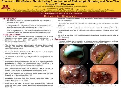
Introduction: Bilo-enteric fistulas are an uncommon complication after placement of biliary drains. Most reports describe surgical management for bilo-enteric fistulas and reports of these fistulas managed endoscopically are rare. Here, we report a case of a bilo-enteric fistula which was managed by combination therapy with endoscopic suturing and over-the-scope-clip.
Case Description/Methods: A 75-year-old man underwent laparoscopic cholecystectomy for acute cholecystitis, which was complicated by injury to the common bile duct and required conversion to an open cholecystectomy and Roux-en-Y hepaticojejunostomy. After discharge, he returned with an ongoing bile leak and interventional radiology (IR) placed an internal-external biliary drain across the hepaticojejunostomy anastomosis. Following IR drainage, he had persistent fever and leukocytosis. Imaging revealed a perihepatic abscess and biloma. He underwent an additional IR-guided percutaneous drain placement into the biloma. Subsequently, cholangiogram revealed bile leak at the hepaticojejunostomy site with extension of contrast into the proximal duodenum concerning for a fistulous communication between the duodenum and biloma.
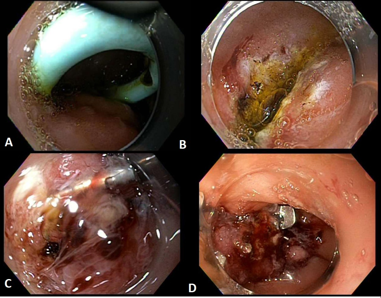
After multidisciplinary discussion, the decision was made to evaluate the fistulous communication with esophagogastroduodenoscopy (EGD). An EGD was performed and the previously placed external drain was seen fistulized into the duodenal bulb [Figure 1A]. The external drain was pulled back outside the duodenal lumen. The fistulous opening was 15 mm in size. Argon plasma coagulation of the tract edges was performed to promote tissue healing and closure followed by endoscopic suturing to close the tract [Figure 1B]. Endoscopic suturing was performed using the OverSitch device and one suture was placed in a figure of eight fashion. However, a 3-mm opening was seen immediately distal to the pylorus even after cinch placement [Figure 1C]. To close the remaining defect, a 12-mm in diameter over-the-scope clip was placed [Figure 1D]. Following closure, there was no residual contrast leakage confirming successful closure of the fistula. The external drain was subsequently removed without evidence of biloma re-accumulation on follow-up imaging.
Discussion: Our report highlights that a combination of endoscopic suturing and over-the-scope clip closure approach can be a potential endoscopic option for managing bilo-enteric fistulas.

Disclosures:
Taiwo Ajose, MD1, Krystal Mills, MD2, Nicholas McDonald, MD3, Idowu Ajose, MBBS3, Bryant Megna, MD3, Mohammad Bilal, MD4. D0290 - Closure of Bilo-Entetric Fistula Using Combination of Endoscopic Suturing and Over-the-Scope Clip Placement, ACG 2022 Annual Scientific Meeting Abstracts. Charlotte, NC: American College of Gastroenterology.
1University of Minnesota Medical School, Minneapolis, MN; 2Morehouse School of Medicine, Atlanta, GA; 3University of Minnesota, Minneapolis, MN; 4University of Minnesota, Minneapolis VA Medical Center, Minneapolis, MN
Introduction: Bilo-enteric fistulas are an uncommon complication after placement of biliary drains. Most reports describe surgical management for bilo-enteric fistulas and reports of these fistulas managed endoscopically are rare. Here, we report a case of a bilo-enteric fistula which was managed by combination therapy with endoscopic suturing and over-the-scope-clip.
Case Description/Methods: A 75-year-old man underwent laparoscopic cholecystectomy for acute cholecystitis, which was complicated by injury to the common bile duct and required conversion to an open cholecystectomy and Roux-en-Y hepaticojejunostomy. After discharge, he returned with an ongoing bile leak and interventional radiology (IR) placed an internal-external biliary drain across the hepaticojejunostomy anastomosis. Following IR drainage, he had persistent fever and leukocytosis. Imaging revealed a perihepatic abscess and biloma. He underwent an additional IR-guided percutaneous drain placement into the biloma. Subsequently, cholangiogram revealed bile leak at the hepaticojejunostomy site with extension of contrast into the proximal duodenum concerning for a fistulous communication between the duodenum and biloma.
After multidisciplinary discussion, the decision was made to evaluate the fistulous communication with esophagogastroduodenoscopy (EGD). An EGD was performed and the previously placed external drain was seen fistulized into the duodenal bulb [Figure 1A]. The external drain was pulled back outside the duodenal lumen. The fistulous opening was 15 mm in size. Argon plasma coagulation of the tract edges was performed to promote tissue healing and closure followed by endoscopic suturing to close the tract [Figure 1B]. Endoscopic suturing was performed using the OverSitch device and one suture was placed in a figure of eight fashion. However, a 3-mm opening was seen immediately distal to the pylorus even after cinch placement [Figure 1C]. To close the remaining defect, a 12-mm in diameter over-the-scope clip was placed [Figure 1D]. Following closure, there was no residual contrast leakage confirming successful closure of the fistula. The external drain was subsequently removed without evidence of biloma re-accumulation on follow-up imaging.
Discussion: Our report highlights that a combination of endoscopic suturing and over-the-scope clip closure approach can be a potential endoscopic option for managing bilo-enteric fistulas.

Figure: Figure 1A- External drain seen fistulized into the duodenal bulb
Figure 1B- Endoscopic suturing to close the fistula tract
Figure 1C- Pylorus opening seen after endoscopic suturing
Figure 1D- 12-mm over-the-scope clip was placed to close the defect
Figure 1B- Endoscopic suturing to close the fistula tract
Figure 1C- Pylorus opening seen after endoscopic suturing
Figure 1D- 12-mm over-the-scope clip was placed to close the defect
Disclosures:
Taiwo Ajose indicated no relevant financial relationships.
Krystal Mills indicated no relevant financial relationships.
Nicholas McDonald indicated no relevant financial relationships.
Idowu Ajose indicated no relevant financial relationships.
Bryant Megna indicated no relevant financial relationships.
Mohammad Bilal indicated no relevant financial relationships.
Taiwo Ajose, MD1, Krystal Mills, MD2, Nicholas McDonald, MD3, Idowu Ajose, MBBS3, Bryant Megna, MD3, Mohammad Bilal, MD4. D0290 - Closure of Bilo-Entetric Fistula Using Combination of Endoscopic Suturing and Over-the-Scope Clip Placement, ACG 2022 Annual Scientific Meeting Abstracts. Charlotte, NC: American College of Gastroenterology.
