Back


Poster Session A - Sunday Afternoon
Category: Esophagus
A0209 - Successful Treatment of a T2 Esophageal Adenocarcinoma With Endoscopic Mucosal Resection
Sunday, October 23, 2022
5:00 PM – 7:00 PM ET
Location: Crown Ballroom

Has Audio

Praneeth Kudaravalli, MD
Augusta University Medical Center
Augusta, GA
Presenting Author(s)
Praneeth Kudaravalli, MD1, Aneesha Gummadi, MS2, Jin Yulan, MD2, Kwabena O. Adu-Gyamfi, MBChB3, Viveksandeep Chandrasekar, MBBS4, Subbaramiah Sridhar, MBBS, MPH5, John Erikson L. Yap, MD4
1Augusta University Medical Center, Augusta, GA; 2Augusta University, Augusta, GA; 3Medical College of Georgia - Augusta University, Augusta, GA; 4Augusta University Medical College of Georiga, Augusta, GA; 5Medical College of Georgia, Augusta, GA
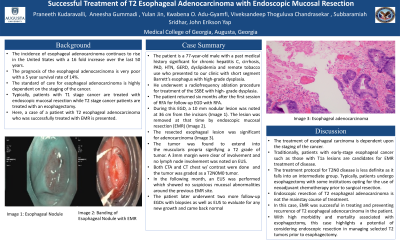
Introduction: The incidence of esophageal adenocarcinoma (EAC) continues to rise in the US with a 16-fold increase over the last 50 years. The prognosis of the disease is very poor with a 5-year survival rate of only 14%. The standard of care for EAC is dependent on the TNM stage of the cancer. Typically, patients with T1a stage cancer are treated with Endoscopic Mucosal Resection (EMR) while T2 stage and above are treated with esophagectomy. We present a patient with nodular BE with dysplasia who was successfully treated with EMR, later identified histologically as T2 stage.
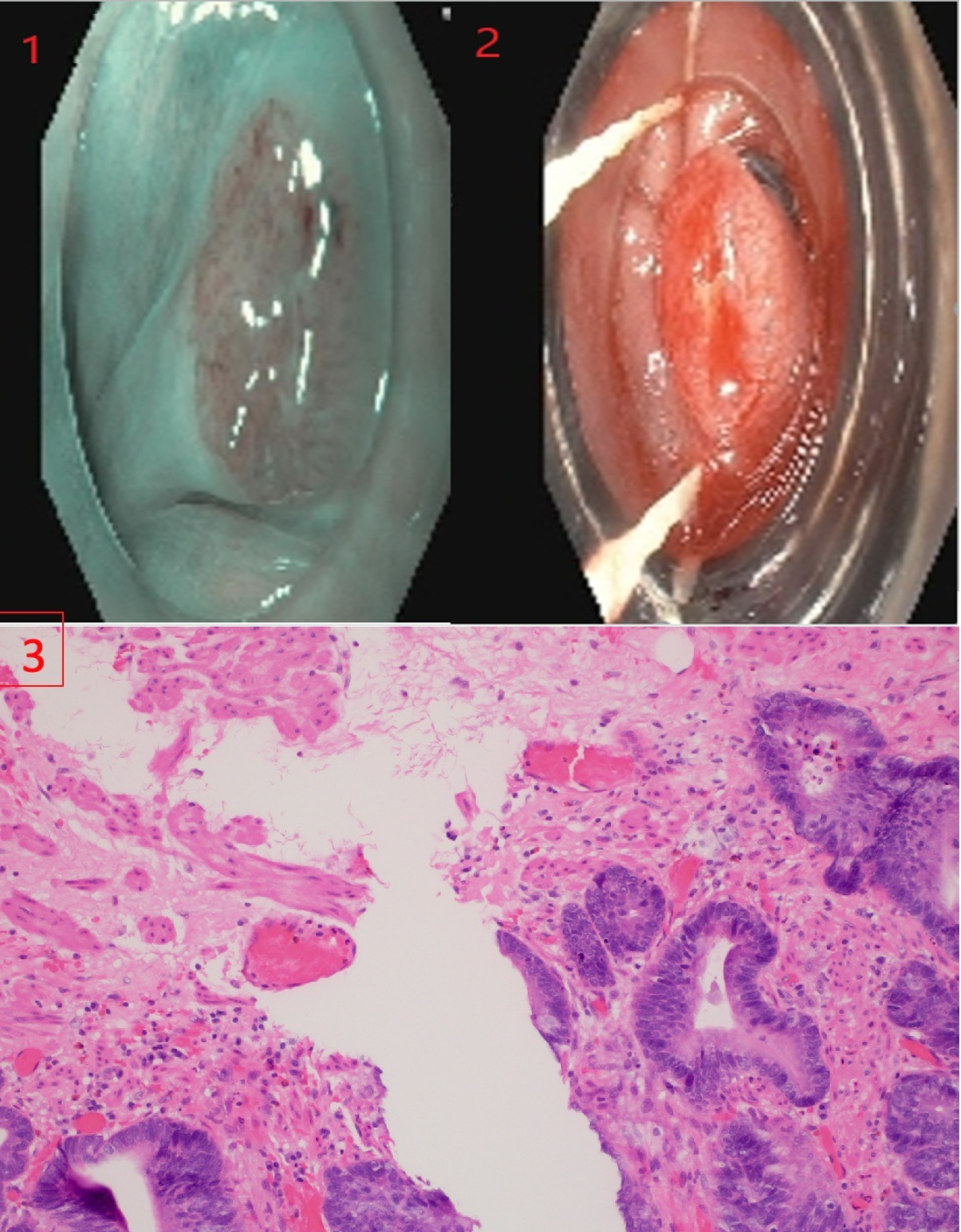
Case Description/Methods: A 77-yr old male with past medical history significant for cirrhosis secondary to chronic hepatitis C, GERD, and remote tobacco use was referred for management of his short segment Barrett’s Esophagus with high grade dysplasia with focal radio frequency ablation (RFA). His vitals and physical exam were unremarkable. 6-months following the initial session of RFA, patient returned for an EGD and was noted to have 10mm nodular lesion in the distal esophagus at 36cm from the incisors (Image 1). The lesion was removed via band-EMR (Image 2). Pathology came back as EAC with the tumor extending into the muscularis propria signifying a T2 grade tumor. All margins and edges (at least 3mm) were uninvolved, and no lymph node involvement was noted. CT abdomen and chest showed no evidence of metastatic disease. The tumor was graded as a T2N0M0. An endoscopic ultrasound (EUS) was performed which showed no suspicious mucosal abnormalities. The patient subsequently underwent 2 more follow up EGDs with biopsies and a EUS to evaluate for evidence of residual lesion. There was no evidence of dysplasia or carcinoma noted on imaging and biopsies.
Discussion: The treatment protocol for T2N0M0 disease is not well defined as it falls into an intermediate group. Typically, patients undergo esophagectomy with some institutions opting to use neoadjuvant chemotherapy prior to surgical resection. Of note, EMR of T2 esophageal adenocarcinoma is not the mainstay of treatment. However, in our case, EMR was successful in treating T2 EAC. With high morbidity and mortality associated with esophagectomy especially in the presence of other co-morbidities like cirrhosis, our case highlights the importance of possibly considering EMR for selected T2 tumors as an alternative option especially in high risk surgical groups.

Disclosures:
Praneeth Kudaravalli, MD1, Aneesha Gummadi, MS2, Jin Yulan, MD2, Kwabena O. Adu-Gyamfi, MBChB3, Viveksandeep Chandrasekar, MBBS4, Subbaramiah Sridhar, MBBS, MPH5, John Erikson L. Yap, MD4. A0209 - Successful Treatment of a T2 Esophageal Adenocarcinoma With Endoscopic Mucosal Resection, ACG 2022 Annual Scientific Meeting Abstracts. Charlotte, NC: American College of Gastroenterology.
1Augusta University Medical Center, Augusta, GA; 2Augusta University, Augusta, GA; 3Medical College of Georgia - Augusta University, Augusta, GA; 4Augusta University Medical College of Georiga, Augusta, GA; 5Medical College of Georgia, Augusta, GA
Introduction: The incidence of esophageal adenocarcinoma (EAC) continues to rise in the US with a 16-fold increase over the last 50 years. The prognosis of the disease is very poor with a 5-year survival rate of only 14%. The standard of care for EAC is dependent on the TNM stage of the cancer. Typically, patients with T1a stage cancer are treated with Endoscopic Mucosal Resection (EMR) while T2 stage and above are treated with esophagectomy. We present a patient with nodular BE with dysplasia who was successfully treated with EMR, later identified histologically as T2 stage.
Case Description/Methods: A 77-yr old male with past medical history significant for cirrhosis secondary to chronic hepatitis C, GERD, and remote tobacco use was referred for management of his short segment Barrett’s Esophagus with high grade dysplasia with focal radio frequency ablation (RFA). His vitals and physical exam were unremarkable. 6-months following the initial session of RFA, patient returned for an EGD and was noted to have 10mm nodular lesion in the distal esophagus at 36cm from the incisors (Image 1). The lesion was removed via band-EMR (Image 2). Pathology came back as EAC with the tumor extending into the muscularis propria signifying a T2 grade tumor. All margins and edges (at least 3mm) were uninvolved, and no lymph node involvement was noted. CT abdomen and chest showed no evidence of metastatic disease. The tumor was graded as a T2N0M0. An endoscopic ultrasound (EUS) was performed which showed no suspicious mucosal abnormalities. The patient subsequently underwent 2 more follow up EGDs with biopsies and a EUS to evaluate for evidence of residual lesion. There was no evidence of dysplasia or carcinoma noted on imaging and biopsies.
Discussion: The treatment protocol for T2N0M0 disease is not well defined as it falls into an intermediate group. Typically, patients undergo esophagectomy with some institutions opting to use neoadjuvant chemotherapy prior to surgical resection. Of note, EMR of T2 esophageal adenocarcinoma is not the mainstay of treatment. However, in our case, EMR was successful in treating T2 EAC. With high morbidity and mortality associated with esophagectomy especially in the presence of other co-morbidities like cirrhosis, our case highlights the importance of possibly considering EMR for selected T2 tumors as an alternative option especially in high risk surgical groups.

Figure: Picture 1 - (1.1): Esophageal Nodule. (1.2): Banding of Esophageal Nodule with EMR. (1.3) Histopathology of esophageal adenocarcinoma.
Disclosures:
Praneeth Kudaravalli indicated no relevant financial relationships.
Aneesha Gummadi indicated no relevant financial relationships.
Jin Yulan indicated no relevant financial relationships.
Kwabena Adu-Gyamfi indicated no relevant financial relationships.
Viveksandeep Chandrasekar indicated no relevant financial relationships.
Subbaramiah Sridhar indicated no relevant financial relationships.
John Erikson Yap indicated no relevant financial relationships.
Praneeth Kudaravalli, MD1, Aneesha Gummadi, MS2, Jin Yulan, MD2, Kwabena O. Adu-Gyamfi, MBChB3, Viveksandeep Chandrasekar, MBBS4, Subbaramiah Sridhar, MBBS, MPH5, John Erikson L. Yap, MD4. A0209 - Successful Treatment of a T2 Esophageal Adenocarcinoma With Endoscopic Mucosal Resection, ACG 2022 Annual Scientific Meeting Abstracts. Charlotte, NC: American College of Gastroenterology.
