Back


Poster Session D - Tuesday Morning
Category: Esophagus
D0246 - Rare Chest Pain Presentation of Esophageal Intramural Pseudodiverticulosis in AIDS Patient
Tuesday, October 25, 2022
10:00 AM – 12:00 PM ET
Location: Crown Ballroom

Has Audio

Lavannya Atri, MS
Augusta University
Augusta, GA
Presenting Author(s)
Praneeth Kudaravalli, MD1, Lavannya Atri, MS2, Dariush Shahsavari, MD3, Viveksandeep Chandrasekar, MBBS4, John Erikson L. Yap, MD4, Zain A. Sobani, MD5
1Augusta University Medical Center, Augusta, GA; 2Augusta University, Augusta, GA; 3Medical College of Georgia - Augusta University, Augusta, GA; 4Augusta University Medical College of Georiga, Augusta, GA; 5Augusta University/Medical College of Georgia, Augusta, GA
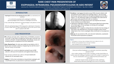
Introduction: Esophageal intramural pseudodiverticulosis (EIP) is an extremely rare condition. It is commonly associated with esophageal candidiasis, gastroesophageal reflux disease or esophageal carcinoma. It has a bimodal age distribution among teenagers and patients in their 50s. With the pathophysiology of EIP being poorly understood, it becomes crucial to make note of unique presentations such as this case of chest pain in a patient with AIDS.
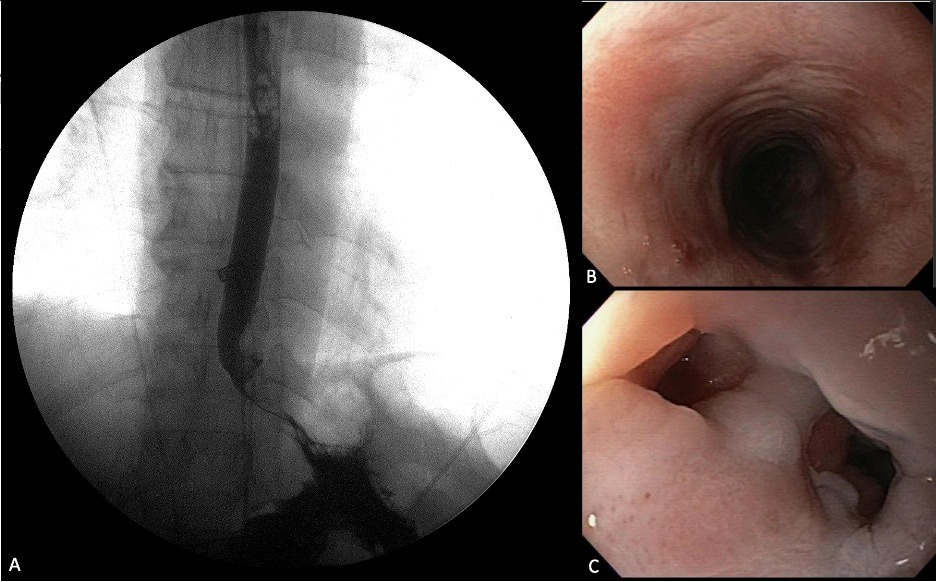
Case Description/Methods: A 58-year-old male with a medical history of HIV/AIDS, presented to ED with a 5-day history of progressive worsening of chest pain and shortness of breath. He denied dysphagia, odynophagia, nausea and vomiting. Patient is nonadherent to his HIV medications. His vitals were stable but was febrile at 39.5 C. Physical exam revealed a cachectic, ill-appearing man with the rest of the exam being unremarkable. Labs revealed an absolute neutrophil count of 800 cells/mm, CD4 count of 53 cells/mm and negative blood cultures. Acute coronary syndrome was ruled out. He was treated with cefepime for his neutropenic fever. CT of the chest and abdomen showed distal esophagitis with a small hiatal hernia, no pneumonia or pulmonary embolism. Esophagogastroduodenoscopy (EGD) showed multiple small pseudodiverticula throughout the esophagus and a large diverticulum noted just proximal to the gastroesophageal junction (GEJ). No endoscopic evidence of esophagitis was noted and the gastroscope easily passed the GEJ with no resistance. The area adjacent to the distal diverticulum was biopsied and showed chronic and focal acute inflammation. A subsequent esophogram showed a small contrast-filled focal outpouching at the distal aspect of the esophagus correlating with esophageal diverticulum. After a negative cardiopulmonary evaluation for the etiology of chest pain it was attributed to an uncommon symptom of EIP. Chronic inflammation from chronic esophagitis likely contributed to the progression of EIP. He was started on PPIs, anti-retroviral medications, prophylactic fluconazole which improved his symptoms.
Discussion: Chest pain without dysphagia is an extremely uncommon presenting symptom of EIP. Once cardiac etiology of chest pain is excluded, an EGD should follow to further assess the etiology of symptoms and if EIP is noted it is important to rule out complications such as a mediastinal fistula formation and perforation. Treatment should include management of the underlying inflammatory/infectious pathology such as HIV in this patient.

Disclosures:
Praneeth Kudaravalli, MD1, Lavannya Atri, MS2, Dariush Shahsavari, MD3, Viveksandeep Chandrasekar, MBBS4, John Erikson L. Yap, MD4, Zain A. Sobani, MD5. D0246 - Rare Chest Pain Presentation of Esophageal Intramural Pseudodiverticulosis in AIDS Patient, ACG 2022 Annual Scientific Meeting Abstracts. Charlotte, NC: American College of Gastroenterology.
1Augusta University Medical Center, Augusta, GA; 2Augusta University, Augusta, GA; 3Medical College of Georgia - Augusta University, Augusta, GA; 4Augusta University Medical College of Georiga, Augusta, GA; 5Augusta University/Medical College of Georgia, Augusta, GA
Introduction: Esophageal intramural pseudodiverticulosis (EIP) is an extremely rare condition. It is commonly associated with esophageal candidiasis, gastroesophageal reflux disease or esophageal carcinoma. It has a bimodal age distribution among teenagers and patients in their 50s. With the pathophysiology of EIP being poorly understood, it becomes crucial to make note of unique presentations such as this case of chest pain in a patient with AIDS.
Case Description/Methods: A 58-year-old male with a medical history of HIV/AIDS, presented to ED with a 5-day history of progressive worsening of chest pain and shortness of breath. He denied dysphagia, odynophagia, nausea and vomiting. Patient is nonadherent to his HIV medications. His vitals were stable but was febrile at 39.5 C. Physical exam revealed a cachectic, ill-appearing man with the rest of the exam being unremarkable. Labs revealed an absolute neutrophil count of 800 cells/mm, CD4 count of 53 cells/mm and negative blood cultures. Acute coronary syndrome was ruled out. He was treated with cefepime for his neutropenic fever. CT of the chest and abdomen showed distal esophagitis with a small hiatal hernia, no pneumonia or pulmonary embolism. Esophagogastroduodenoscopy (EGD) showed multiple small pseudodiverticula throughout the esophagus and a large diverticulum noted just proximal to the gastroesophageal junction (GEJ). No endoscopic evidence of esophagitis was noted and the gastroscope easily passed the GEJ with no resistance. The area adjacent to the distal diverticulum was biopsied and showed chronic and focal acute inflammation. A subsequent esophogram showed a small contrast-filled focal outpouching at the distal aspect of the esophagus correlating with esophageal diverticulum. After a negative cardiopulmonary evaluation for the etiology of chest pain it was attributed to an uncommon symptom of EIP. Chronic inflammation from chronic esophagitis likely contributed to the progression of EIP. He was started on PPIs, anti-retroviral medications, prophylactic fluconazole which improved his symptoms.
Discussion: Chest pain without dysphagia is an extremely uncommon presenting symptom of EIP. Once cardiac etiology of chest pain is excluded, an EGD should follow to further assess the etiology of symptoms and if EIP is noted it is important to rule out complications such as a mediastinal fistula formation and perforation. Treatment should include management of the underlying inflammatory/infectious pathology such as HIV in this patient.

Figure: Figure 1. A. Esophagogram showing a large esophageal diverticulum adjacent to the LES. B. Multiple small pseudodiverticula in the upper esophagus. C. Large esophageal diverticula adjacent to LES
Disclosures:
Praneeth Kudaravalli indicated no relevant financial relationships.
Lavannya Atri indicated no relevant financial relationships.
Dariush Shahsavari indicated no relevant financial relationships.
Viveksandeep Chandrasekar indicated no relevant financial relationships.
John Erikson Yap indicated no relevant financial relationships.
Zain Sobani indicated no relevant financial relationships.
Praneeth Kudaravalli, MD1, Lavannya Atri, MS2, Dariush Shahsavari, MD3, Viveksandeep Chandrasekar, MBBS4, John Erikson L. Yap, MD4, Zain A. Sobani, MD5. D0246 - Rare Chest Pain Presentation of Esophageal Intramural Pseudodiverticulosis in AIDS Patient, ACG 2022 Annual Scientific Meeting Abstracts. Charlotte, NC: American College of Gastroenterology.
