Back


Poster Session E - Tuesday Afternoon
Category: Endoscopy Video Forum
E0184 - Pancreatoscopy Using Adult Gastroscope as a Diagnostic Method for Main Duct IPMN
Tuesday, October 25, 2022
3:00 PM – 5:00 PM ET
Location: Crown Ballroom

Has Audio
- GK
Grace E. Kim, MD
The University of Chicago Medical Center
Chicago, Illinois
Presenting Author(s)
Award: Presidential Poster Award
Grace E. Kim, MD1, David Y. Lo, MD, FACG2
1The University of Chicago Medical Center, Chicago, IL; 2Ohio Gastroenterology Group, Inc, and The Ohio State University College of Medicine, Columbus, OH
Introduction: Intraductal papillary mucinous neoplasm (IPMN) is a premalignant condition of the pancreas characterized by epithelial neoplasms of the pancreatic duct. Typically, IPMNs are diagnosed by cross sectional imaging and/or endoscopic ultrasound owing to its high sensitivity. Less commonly, pancreatoscopy is utilized for direct visualization of the pancreatic duct using a single operator cholangioscope but it has limitations. There are case reports of using thinner gastroscopes to achieve pancreatoscopy, however to date, there are no reports of using adult gastroscopes to directly visualize the main pancreatic duct.
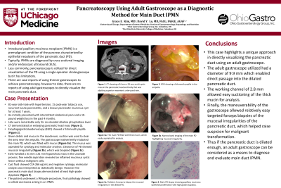
Case Description/Methods: We present an 81-year-old-male with hypertension, 15 pack-year tobacco use, recurrent acute pancreatitis, and a known pancreatic mucinous cyst for at least 7 years. He initially presented with intermittent abdominal pain and a 10-pound weight loss in the past 4 months. Labs were remarkable only for an elevated alkaline phosphatase level. He underwent CT that showed an enlarging pancreatic head mass. The patient underwent esophagogastroduodenoscopy (EGD) which showed a fishmouth papilla in the ampulla. Due to the presence of debris and mucus in the duodenum, suction was used to clear the area near the ampulla, however the gastroscope inadvertently intubated the main pancreatic duct. Endoscopic ultrasound was then performed which showed an oval, hypoechoic mass measuring 31 mm x 21 mm in the uncinate process. This was aspirated via fine needle aspiration. Interestingly, the molecular analysis was interpreted as statistically benign; however the pancreatic main duct biopsy showed a superficial portion of ductal wall with acute and chronic inflammation, papillary mucinous epithelial proliferation, and focal marked epithelial atypia consistent with at least high-grade dysplasia.
Discussion: This case highlights a unique approach in directly visualizing the pancreatic duct using an adult gastroscope. The adult gastroscope used in this case had a diameter of 9.9mm which enabled direct passage into the dilated pancreatic duct. It also had a working channel of 2.8mm which allowed easy suctioning of the thick mucin for analysis. Finally, the maneuverability of the gastroscope allowed relatively easy targeted forceps biopsies of the mucosal irregularities of the pancreatic duct, which helped raise suspicion for malignant transformation. Thus if the pancreatic duct is dilated enough, an adult gastroscope can be considered as a means to diagnose main duct IPMN.
Disclosures:
Grace E. Kim, MD1, David Y. Lo, MD, FACG2. E0184 - Pancreatoscopy Using Adult Gastroscope as a Diagnostic Method for Main Duct IPMN, ACG 2022 Annual Scientific Meeting Abstracts. Charlotte, NC: American College of Gastroenterology.
Grace E. Kim, MD1, David Y. Lo, MD, FACG2
1The University of Chicago Medical Center, Chicago, IL; 2Ohio Gastroenterology Group, Inc, and The Ohio State University College of Medicine, Columbus, OH
Introduction: Intraductal papillary mucinous neoplasm (IPMN) is a premalignant condition of the pancreas characterized by epithelial neoplasms of the pancreatic duct. Typically, IPMNs are diagnosed by cross sectional imaging and/or endoscopic ultrasound owing to its high sensitivity. Less commonly, pancreatoscopy is utilized for direct visualization of the pancreatic duct using a single operator cholangioscope but it has limitations. There are case reports of using thinner gastroscopes to achieve pancreatoscopy, however to date, there are no reports of using adult gastroscopes to directly visualize the main pancreatic duct.
Case Description/Methods: We present an 81-year-old-male with hypertension, 15 pack-year tobacco use, recurrent acute pancreatitis, and a known pancreatic mucinous cyst for at least 7 years. He initially presented with intermittent abdominal pain and a 10-pound weight loss in the past 4 months. Labs were remarkable only for an elevated alkaline phosphatase level. He underwent CT that showed an enlarging pancreatic head mass. The patient underwent esophagogastroduodenoscopy (EGD) which showed a fishmouth papilla in the ampulla. Due to the presence of debris and mucus in the duodenum, suction was used to clear the area near the ampulla, however the gastroscope inadvertently intubated the main pancreatic duct. Endoscopic ultrasound was then performed which showed an oval, hypoechoic mass measuring 31 mm x 21 mm in the uncinate process. This was aspirated via fine needle aspiration. Interestingly, the molecular analysis was interpreted as statistically benign; however the pancreatic main duct biopsy showed a superficial portion of ductal wall with acute and chronic inflammation, papillary mucinous epithelial proliferation, and focal marked epithelial atypia consistent with at least high-grade dysplasia.
Discussion: This case highlights a unique approach in directly visualizing the pancreatic duct using an adult gastroscope. The adult gastroscope used in this case had a diameter of 9.9mm which enabled direct passage into the dilated pancreatic duct. It also had a working channel of 2.8mm which allowed easy suctioning of the thick mucin for analysis. Finally, the maneuverability of the gastroscope allowed relatively easy targeted forceps biopsies of the mucosal irregularities of the pancreatic duct, which helped raise suspicion for malignant transformation. Thus if the pancreatic duct is dilated enough, an adult gastroscope can be considered as a means to diagnose main duct IPMN.
Disclosures:
Grace Kim indicated no relevant financial relationships.
David Lo indicated no relevant financial relationships.
Grace E. Kim, MD1, David Y. Lo, MD, FACG2. E0184 - Pancreatoscopy Using Adult Gastroscope as a Diagnostic Method for Main Duct IPMN, ACG 2022 Annual Scientific Meeting Abstracts. Charlotte, NC: American College of Gastroenterology.

