Back


Poster Session C - Monday Afternoon
Category: Biliary/Pancreas
C0063 - Rare Presentations of Metastatic Urothelial Carcinoma to GI Tract: A Case Series Highlighting Key Principles of Endoscopy in the Cancer Patient
Monday, October 24, 2022
3:00 PM – 5:00 PM ET
Location: Crown Ballroom

Has Audio

Timothy Lee, MD
University of Rochester Medical Center
Rochester, NY
Presenting Author(s)
Timothy Lee, MD, Amulya Penmetsa, MD, Jonathan Huang, DO, Asad Ullah, MD, Sarah Enslin, PA-C, Vivek Kaul, MD, FACG
University of Rochester Medical Center, Rochester, NY
Introduction: In the US, ~80,000 new cases of bladder cancer are diagnosed yearly, 4% of which present with metastatic disease (lymph nodes, liver, peritoneum, lung). Most bladder cancers are pure urothelial carcinoma (Uca). Metastatic spread to the biliary system and small bowel has been rarely described. We present a report of 3 patients with known urothelial carcinoma who presented with gastrointestinal (GI) metastasis.
Case Description/Methods: Case 1
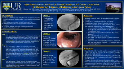
A 71-year-old male with Uca presented with cholangitis. Endoscopic retrograde cholangiopancreatography (ERCP) was done, subhilar biliary stricture found, brush cytology performed, biliary stent placed (Fig.1). Endoscopic ultrasonography (EUS) revealed an ill-defined hypoechoic peribiliary mass. Fine-needle aspiration (FNA) cytology confirmed malignant cells from metastatic Uca.
Case 2
A 63-year-old male with UCa presented with gastric outlet obstruction (GOO). Imaging revealed a possible pancreatic head mass. Biopsies of the duodenum were non-diagnostic initially. EUS-FNA of the pancreatic head mass revealed benign pancreatic cells. Endoscopy was repeated with extensive biopsies taken of a lumen obstructing externally infiltrating duodenal mass/stricture (Fig.2) which confirmed poorly differentiated carcinoma consistent with metastatic Uca. A duodenal stent was placed.
Case 3
A 68-year-old male with Uca with metastases to the liver and omentum presented with abdominal pain and biliary obstruction. ERCP was performed with biliary stent placement across the biliary stricture. Brush cytology revealed malignant cells of urothelial origin, representative of metastasis to the CBD
Discussion: Metastatic Uca to the biliary tree and duodenum has been rarely reported. GOO from Uca is also unusual but may occur due to retroperitoneal, hepatic flexure or duodenal masses. Endoscopic evaluation can be challenging due to altered anatomy. This case series emphasizes several important points:
< !1. In the appropriate clinical context, non-GI malignancies should be in the differential diagnosis when managing biliary obstruction and GOO
< !2.Role of repeat endoluminal biopsy if initial effort is non-diagnostic
< !3.Role of EUS-FNA for tissue sampling
< !4. Role of ERCP and biliary stenting for palliation of biliary obstruction
< !5. Importance of an accurate pathologic diagnosis in a patient with suspected metastatic malignancy, given management and prognostic implications

Disclosures:
Timothy Lee, MD, Amulya Penmetsa, MD, Jonathan Huang, DO, Asad Ullah, MD, Sarah Enslin, PA-C, Vivek Kaul, MD, FACG. C0063 - Rare Presentations of Metastatic Urothelial Carcinoma to GI Tract: A Case Series Highlighting Key Principles of Endoscopy in the Cancer Patient, ACG 2022 Annual Scientific Meeting Abstracts. Charlotte, NC: American College of Gastroenterology.
University of Rochester Medical Center, Rochester, NY
Introduction: In the US, ~80,000 new cases of bladder cancer are diagnosed yearly, 4% of which present with metastatic disease (lymph nodes, liver, peritoneum, lung). Most bladder cancers are pure urothelial carcinoma (Uca). Metastatic spread to the biliary system and small bowel has been rarely described. We present a report of 3 patients with known urothelial carcinoma who presented with gastrointestinal (GI) metastasis.
Case Description/Methods: Case 1
A 71-year-old male with Uca presented with cholangitis. Endoscopic retrograde cholangiopancreatography (ERCP) was done, subhilar biliary stricture found, brush cytology performed, biliary stent placed (Fig.1). Endoscopic ultrasonography (EUS) revealed an ill-defined hypoechoic peribiliary mass. Fine-needle aspiration (FNA) cytology confirmed malignant cells from metastatic Uca.
Case 2
A 63-year-old male with UCa presented with gastric outlet obstruction (GOO). Imaging revealed a possible pancreatic head mass. Biopsies of the duodenum were non-diagnostic initially. EUS-FNA of the pancreatic head mass revealed benign pancreatic cells. Endoscopy was repeated with extensive biopsies taken of a lumen obstructing externally infiltrating duodenal mass/stricture (Fig.2) which confirmed poorly differentiated carcinoma consistent with metastatic Uca. A duodenal stent was placed.
Case 3
A 68-year-old male with Uca with metastases to the liver and omentum presented with abdominal pain and biliary obstruction. ERCP was performed with biliary stent placement across the biliary stricture. Brush cytology revealed malignant cells of urothelial origin, representative of metastasis to the CBD
Discussion: Metastatic Uca to the biliary tree and duodenum has been rarely reported. GOO from Uca is also unusual but may occur due to retroperitoneal, hepatic flexure or duodenal masses. Endoscopic evaluation can be challenging due to altered anatomy. This case series emphasizes several important points:
< !1. In the appropriate clinical context, non-GI malignancies should be in the differential diagnosis when managing biliary obstruction and GOO
< !2.Role of repeat endoluminal biopsy if initial effort is non-diagnostic
< !3.Role of EUS-FNA for tissue sampling
< !4. Role of ERCP and biliary stenting for palliation of biliary obstruction
< !5. Importance of an accurate pathologic diagnosis in a patient with suspected metastatic malignancy, given management and prognostic implications

Figure: Fig 1a: Cholangiogram from ERCP for Case 1 demonstrating a high-grade subhilar biliary stricture
Fig 1b: Infiltrating, obstructing duodenal mass with stricture secondary to urothelial carcinoma metastases in Case 2
Fig 1c: Cholangiogram from ERCP for Case 3 demonstrating a long stricture of the distal CBD and the mid CBD
Fig 1b: Infiltrating, obstructing duodenal mass with stricture secondary to urothelial carcinoma metastases in Case 2
Fig 1c: Cholangiogram from ERCP for Case 3 demonstrating a long stricture of the distal CBD and the mid CBD
Disclosures:
Timothy Lee indicated no relevant financial relationships.
Amulya Penmetsa indicated no relevant financial relationships.
Jonathan Huang indicated no relevant financial relationships.
Asad Ullah indicated no relevant financial relationships.
Sarah Enslin indicated no relevant financial relationships.
Vivek Kaul: Ambu – Consultant. CDX – Consultant. Cook Medical – Consultant. Motus GI – Consultant. Steris Corp – Consultant.
Timothy Lee, MD, Amulya Penmetsa, MD, Jonathan Huang, DO, Asad Ullah, MD, Sarah Enslin, PA-C, Vivek Kaul, MD, FACG. C0063 - Rare Presentations of Metastatic Urothelial Carcinoma to GI Tract: A Case Series Highlighting Key Principles of Endoscopy in the Cancer Patient, ACG 2022 Annual Scientific Meeting Abstracts. Charlotte, NC: American College of Gastroenterology.

