Back


Poster Session E - Tuesday Afternoon
Category: Esophagus
E0251 - Multifocal Pyloric Gland Adenoma of the Esophagus Treated by Circumferential Endoscopic Submucosal Dissection
Tuesday, October 25, 2022
3:00 PM – 5:00 PM ET
Location: Crown Ballroom

Has Audio

Howard Lee, MD
San Antonio Uniformed Services Health Education Consortium
SAN ANTONIO, TX
Presenting Author(s)
Award: Presidential Poster Award
Howard Lee, MD1, John Magulick, MD2, Carl Kay, MD1, John G. Quiles, MD2
1San Antonio Uniformed Services Health Education Consortium, Fort Sam Houston, TX; 2San Antonio Uniformed Services Health Education Consortium, San Antonio, TX
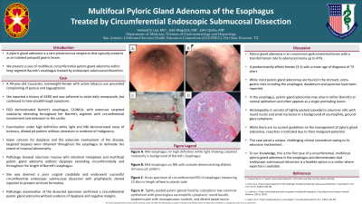
Introduction: A pyloric gland adenoma (PGA) is a rare precancerous neoplasm typically seen as an isolated polypoid lesion in the stomach. We present a case of multifocal, circumferential PGA within long-segment Barrett’s esophagus treated by endoscopic submucosal dissection (ESD).
Case Description/Methods: A 49-year-old female presents complaining of pyrosis and regurgitation. She reports a long history of GERD and adheres to esomeprazole twice daily, but continues to have breakthrough symptoms. Her risk factors include White race, active tobacco use and obesity.
EGD demonstrated endoscopic evidence of Barrett’s esophagus, C10M14, with extensive carpeted nodularity from lateral to posterior wall with circumferential involvement at the GEJ with extension to the cardia. Close examination under high definition white light and NBI showed scattered areas of tortuous, dilated pit pattern without ulceration or evidence of malignancy.
Random and targeted biopsies were obtained throughout the esophagus to demarcate the lateral and circumferential extent of mucosal abnormality and to evaluate for dysplasia. Pathology showed columnar mucosa with intestinal metaplasia and multifocal PGA without dysplasia. Deemed to be a poor candidate for esophagectomy, the patient underwent successful circumferential ESD with prophylactic steroid injection to prevent stricture formation.
Discussion: PGA is an uncommon lesion of the gastrointestinal tract with a transformation rate to adenocarcinoma up to 47%. It predominantly affects female (3:1) with a mean age of diagnosis of 73 years. While most PGAs are observed in the stomach, extragastric sites including the esophagus, duodenum and pancreas have been reported. In the esophagus, PGA may arise in either Barrett’s or normal epithelium and often appears as a single protruding lesion. Histologically, it consists of tightly packed pyloric glands lined by cuboidal to columnar cells with round nuclei and small nucleolus in a background of eosinophilic, ground-glass cytoplasm.
While there is no guideline on the management of PGA, resection is indicated due to its malignant potential. This case poses a unique, challenging clinical conundrum owing to its extensive involvement. To our knowledge, this is the first case of a circumferential, multifocal PGA in the esophagus and we demonstrated ESD to be a feasible management option for select patients in whom close interval repeat EGDs can be performed for both post-procedural complications and recurrence of lesion surveillance.

Disclosures:
Howard Lee, MD1, John Magulick, MD2, Carl Kay, MD1, John G. Quiles, MD2. E0251 - Multifocal Pyloric Gland Adenoma of the Esophagus Treated by Circumferential Endoscopic Submucosal Dissection, ACG 2022 Annual Scientific Meeting Abstracts. Charlotte, NC: American College of Gastroenterology.
Howard Lee, MD1, John Magulick, MD2, Carl Kay, MD1, John G. Quiles, MD2
1San Antonio Uniformed Services Health Education Consortium, Fort Sam Houston, TX; 2San Antonio Uniformed Services Health Education Consortium, San Antonio, TX
Introduction: A pyloric gland adenoma (PGA) is a rare precancerous neoplasm typically seen as an isolated polypoid lesion in the stomach. We present a case of multifocal, circumferential PGA within long-segment Barrett’s esophagus treated by endoscopic submucosal dissection (ESD).
Case Description/Methods: A 49-year-old female presents complaining of pyrosis and regurgitation. She reports a long history of GERD and adheres to esomeprazole twice daily, but continues to have breakthrough symptoms. Her risk factors include White race, active tobacco use and obesity.
EGD demonstrated endoscopic evidence of Barrett’s esophagus, C10M14, with extensive carpeted nodularity from lateral to posterior wall with circumferential involvement at the GEJ with extension to the cardia. Close examination under high definition white light and NBI showed scattered areas of tortuous, dilated pit pattern without ulceration or evidence of malignancy.
Random and targeted biopsies were obtained throughout the esophagus to demarcate the lateral and circumferential extent of mucosal abnormality and to evaluate for dysplasia. Pathology showed columnar mucosa with intestinal metaplasia and multifocal PGA without dysplasia. Deemed to be a poor candidate for esophagectomy, the patient underwent successful circumferential ESD with prophylactic steroid injection to prevent stricture formation.
Discussion: PGA is an uncommon lesion of the gastrointestinal tract with a transformation rate to adenocarcinoma up to 47%. It predominantly affects female (3:1) with a mean age of diagnosis of 73 years. While most PGAs are observed in the stomach, extragastric sites including the esophagus, duodenum and pancreas have been reported. In the esophagus, PGA may arise in either Barrett’s or normal epithelium and often appears as a single protruding lesion. Histologically, it consists of tightly packed pyloric glands lined by cuboidal to columnar cells with round nuclei and small nucleolus in a background of eosinophilic, ground-glass cytoplasm.
While there is no guideline on the management of PGA, resection is indicated due to its malignant potential. This case poses a unique, challenging clinical conundrum owing to its extensive involvement. To our knowledge, this is the first case of a circumferential, multifocal PGA in the esophagus and we demonstrated ESD to be a feasible management option for select patients in whom close interval repeat EGDs can be performed for both post-procedural complications and recurrence of lesion surveillance.

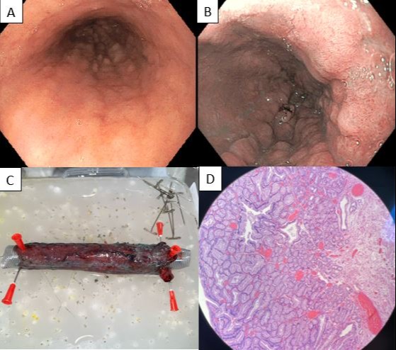
Figure: A. Mid-esophagus on high definition white light showing carpeted nodularity in background of Barrett's Esophagus
B. Mid-esophagus on NBI with nodules demonstrating dilated, tortuous pit pattern
C. Gross specimen of circumferential ESD of esophagus measuring 13.9cm in length affixed to plastic tube.
D. Tightly packed pyloric glands lined by cuboidal or low columnar epithelium with ground glass eosinophilic cytoplasm, round basally located nuclei with inconspicuous nucleoli, and absent apical mucin.
Disclosures:
Howard Lee indicated no relevant financial relationships.
John Magulick indicated no relevant financial relationships.
Carl Kay indicated no relevant financial relationships.
John Quiles indicated no relevant financial relationships.
Howard Lee, MD1, John Magulick, MD2, Carl Kay, MD1, John G. Quiles, MD2. E0251 - Multifocal Pyloric Gland Adenoma of the Esophagus Treated by Circumferential Endoscopic Submucosal Dissection, ACG 2022 Annual Scientific Meeting Abstracts. Charlotte, NC: American College of Gastroenterology.


