Back


Poster Session E - Tuesday Afternoon
Category: GI Bleeding
E0324 - A Rare Case of Simultaneous Bleeding From a Dieulafoy’s Lesion and a Gastroesophageal Varix Type 1 in a Decompensated Cirrhotic Patient
Tuesday, October 25, 2022
3:00 PM – 5:00 PM ET
Location: Crown Ballroom

Has Audio
- NN
Nghia Nguyen, DO
University of Texas Rio Grande Valley at Doctors Hospital at Renaissance
Pharr, Texas
Presenting Author(s)
Nghia Nguyen, DO1, Prateek S. Harne, MBBS, MD2, Asif Zamir, MD, FACG2, Murthy Badiga, MD, FACG2
1University of Texas Rio Grande Valley at Doctors Hospital at Renaissance, Pharr, TX; 2University of Texas Rio Grande Valley at Doctors Hospital at Renaissance, Edinburg, TX
Introduction: Bleeding from Dieulafoy’s lesion (DL) is an important cause of gastrointestinal bleed (GIB) with relatively high mortality. Their incidence is reported to be 1% to 2% of all GIBs1. DLs are most often linked to cardiopulmonary and renal disease2. Bleeding gastro-esophageal varices (GOV) type 1 also pose a high mortality rate (30%). We present a rare case of simultaneous bleeding from an antral DL and a GOV type 1 in a decompensated cirrhotic patient.
Case Description/Methods: A 57-year-old man with a history of decompensated liver cirrhosis complicated by esophageal varices presented with multiple episodes of hematemesis. He denied diarrhea or bloody stool. As per outside records, his most recent EGD 7 months prior was notable for bleeding esophageal varices requiring 3 band ligations. Patient was tachycardic on presentation. Initial labs showed WBC 17.3, Hb 10.7, Hct 34, platelet 166, INR 1.90, Na 132, Cr 0.9, AST 39, ALT 20, ALP 129, total bilirubin 2.9. The patient received blood transfusion and was started on octreotide and pantoprazole infusions. Subsequent EGD revealed a bleeding GOV type 1 treated with three band ligations and interestingly, also a bleeding antral DL which was treated with 3cc epinephrine injection followed by application of three hemoclips. The patient’s clinical status improved with complete resolution of GIB.
Discussion: DL is a rare cause of GIB in advanced liver disease (ALD), which is not directly related to portal hypertension3. It is an abnormally large and tortuous submucosal artery that can rupture and cause life-threatening GIB. With the shift from surgical treatment toward endoscopic interventions, the mortality and morbidity of DLs reportedly improve from 80% to 8.6%1, although, a combined mortality rate from a bleeding DL and GOV remains very high. Endoscopic therapies include epinephrine injection, probe coagulation, band ligation and hemoclips. Mechanical modalities with banding and hemoclips are safest and most effective with lower re-bleeding rate. It was interesting to note that our case had two simultaneous sources of bleeding in the form of GOV type 1 and a DL which were promptly treated. We highlight the importance of thorough endoscopic evaluation when investigating GIBs in ALDs since uncommon causes such as DL can lead to devastating outcome if not being promptly diagnosed and treated.

Disclosures:
Nghia Nguyen, DO1, Prateek S. Harne, MBBS, MD2, Asif Zamir, MD, FACG2, Murthy Badiga, MD, FACG2. E0324 - A Rare Case of Simultaneous Bleeding From a Dieulafoy’s Lesion and a Gastroesophageal Varix Type 1 in a Decompensated Cirrhotic Patient, ACG 2022 Annual Scientific Meeting Abstracts. Charlotte, NC: American College of Gastroenterology.
1University of Texas Rio Grande Valley at Doctors Hospital at Renaissance, Pharr, TX; 2University of Texas Rio Grande Valley at Doctors Hospital at Renaissance, Edinburg, TX
Introduction: Bleeding from Dieulafoy’s lesion (DL) is an important cause of gastrointestinal bleed (GIB) with relatively high mortality. Their incidence is reported to be 1% to 2% of all GIBs1. DLs are most often linked to cardiopulmonary and renal disease2. Bleeding gastro-esophageal varices (GOV) type 1 also pose a high mortality rate (30%). We present a rare case of simultaneous bleeding from an antral DL and a GOV type 1 in a decompensated cirrhotic patient.
Case Description/Methods: A 57-year-old man with a history of decompensated liver cirrhosis complicated by esophageal varices presented with multiple episodes of hematemesis. He denied diarrhea or bloody stool. As per outside records, his most recent EGD 7 months prior was notable for bleeding esophageal varices requiring 3 band ligations. Patient was tachycardic on presentation. Initial labs showed WBC 17.3, Hb 10.7, Hct 34, platelet 166, INR 1.90, Na 132, Cr 0.9, AST 39, ALT 20, ALP 129, total bilirubin 2.9. The patient received blood transfusion and was started on octreotide and pantoprazole infusions. Subsequent EGD revealed a bleeding GOV type 1 treated with three band ligations and interestingly, also a bleeding antral DL which was treated with 3cc epinephrine injection followed by application of three hemoclips. The patient’s clinical status improved with complete resolution of GIB.
Discussion: DL is a rare cause of GIB in advanced liver disease (ALD), which is not directly related to portal hypertension3. It is an abnormally large and tortuous submucosal artery that can rupture and cause life-threatening GIB. With the shift from surgical treatment toward endoscopic interventions, the mortality and morbidity of DLs reportedly improve from 80% to 8.6%1, although, a combined mortality rate from a bleeding DL and GOV remains very high. Endoscopic therapies include epinephrine injection, probe coagulation, band ligation and hemoclips. Mechanical modalities with banding and hemoclips are safest and most effective with lower re-bleeding rate. It was interesting to note that our case had two simultaneous sources of bleeding in the form of GOV type 1 and a DL which were promptly treated. We highlight the importance of thorough endoscopic evaluation when investigating GIBs in ALDs since uncommon causes such as DL can lead to devastating outcome if not being promptly diagnosed and treated.

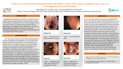
Figure: a. Large amounts of blood noted in the fundus
b. Antral Dieulafoy’s lesion
c. Dieulafoy’s lesion status post banding and epinephrine injection
d. Gastro-esophageal Varix type 1 status post banding
b. Antral Dieulafoy’s lesion
c. Dieulafoy’s lesion status post banding and epinephrine injection
d. Gastro-esophageal Varix type 1 status post banding
Disclosures:
Nghia Nguyen indicated no relevant financial relationships.
Prateek Harne indicated no relevant financial relationships.
Asif Zamir indicated no relevant financial relationships.
Murthy Badiga indicated no relevant financial relationships.
Nghia Nguyen, DO1, Prateek S. Harne, MBBS, MD2, Asif Zamir, MD, FACG2, Murthy Badiga, MD, FACG2. E0324 - A Rare Case of Simultaneous Bleeding From a Dieulafoy’s Lesion and a Gastroesophageal Varix Type 1 in a Decompensated Cirrhotic Patient, ACG 2022 Annual Scientific Meeting Abstracts. Charlotte, NC: American College of Gastroenterology.
