Back
Poster Session E - Tuesday Afternoon
Category: Interventional Endoscopy
E0461 - Combined Antegrade Retrograde Dilation of Radiation-Induced Benign Esophageal Obstruction With Septum Formation: A Case Report
Tuesday, October 25, 2022
3:00 PM – 5:00 PM ET
Location: Crown Ballroom
- WS
Wasef Sayeh, MD
University of Toledo
Toledo, Ohio
Presenting Author(s)
Award: Presidential Poster Award
Wasef Sayeh, MD1, Albert W Tsang, MD2, Jordan Burlen, MD3, Sami Ghazaleh, MD1, Azizullah A. Beran, MD4, Tarik Alhmoud, MD2, Muhannad Heif, MD2
1University of Toledo, Toledo, OH; 2Toledo Hospital, Toledo, OH; 3The Ohio State University, Toledo, OH; 4The University of Toledo, Toledo, OH
Introduction: Approximately 0.8% to 2.6% of patients develop esophageal stenosis and strictures after exposure to radiation therapy for oropharyngeal cancer treatment. Partial esophageal stenosis is usually treated with antegrade dilation. However, this is not possible with complete esophageal strictures. We present a case with radiation induced complete esophageal obstruction with septum formation that was treated using a combined antegrade retrograde dilation (CARD) technique with the use of a needle knife for septotomy.
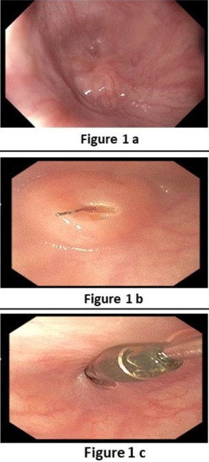
Case Description/Methods: A 76-year-old male patient who was recently diagnosed with stage 3 squamous cell carcinoma at the base of the tongue and has a feeding percutaneous gastrostomy (PEG) tube presented with symptoms of dysphagia and recurrent aspirations three weeks after he completed the last radiation therapy session. A video swallow test was done and showed findings suggestive of esophageal obstruction. Endoscopic evaluation was performed and esophagoscopy demonstrated a benign appearing complete esophageal septum. The scope could not traverse the septum (Figure 1a). Another trial using the Utraslim scope also failed. Then, the CARD technique was performed where an Ultraslim gastroscope was introduced through the PEG tube track and advanced to the level of obstruction in the proximal esophagus. Simultaneously, the adult gastroscope was introduced through the mouth until it reached the level of obstruction. The transillumination from the adult gastroscope was seen by the Ultraslim scope on the opposite side. Free needle septotomy was performed under the guidance of both the transillumination and direct visualization (Figure 1b). A 0.035-inch Jagwire was advanced through the needle knife to the distal side of the esophagus. A Savary wire was advanced under the direct vision of the adult gastroscope through the septotomy. Dilation was performed with a Savary dilator under fluoroscopy and endoscopy guidance with the Ultraslim scope situated in the distal esophagus (Figure 1c). EGD was repeated after two weeks and showed a patent lumen.
Discussion: Complete esophageal stenosis after radiation therapy is not uncommon and should always be kept in mind when patients present with dysphagia symptoms. CARD technique is considered the preferred initial intervention in most cases where a glidewire or suction septotomy are usually used. However, the use of needle knife for septotomy can be considered in selected cases such as in our case.

Disclosures:
Wasef Sayeh, MD1, Albert W Tsang, MD2, Jordan Burlen, MD3, Sami Ghazaleh, MD1, Azizullah A. Beran, MD4, Tarik Alhmoud, MD2, Muhannad Heif, MD2. E0461 - Combined Antegrade Retrograde Dilation of Radiation-Induced Benign Esophageal Obstruction With Septum Formation: A Case Report, ACG 2022 Annual Scientific Meeting Abstracts. Charlotte, NC: American College of Gastroenterology.
Wasef Sayeh, MD1, Albert W Tsang, MD2, Jordan Burlen, MD3, Sami Ghazaleh, MD1, Azizullah A. Beran, MD4, Tarik Alhmoud, MD2, Muhannad Heif, MD2
1University of Toledo, Toledo, OH; 2Toledo Hospital, Toledo, OH; 3The Ohio State University, Toledo, OH; 4The University of Toledo, Toledo, OH
Introduction: Approximately 0.8% to 2.6% of patients develop esophageal stenosis and strictures after exposure to radiation therapy for oropharyngeal cancer treatment. Partial esophageal stenosis is usually treated with antegrade dilation. However, this is not possible with complete esophageal strictures. We present a case with radiation induced complete esophageal obstruction with septum formation that was treated using a combined antegrade retrograde dilation (CARD) technique with the use of a needle knife for septotomy.
Case Description/Methods: A 76-year-old male patient who was recently diagnosed with stage 3 squamous cell carcinoma at the base of the tongue and has a feeding percutaneous gastrostomy (PEG) tube presented with symptoms of dysphagia and recurrent aspirations three weeks after he completed the last radiation therapy session. A video swallow test was done and showed findings suggestive of esophageal obstruction. Endoscopic evaluation was performed and esophagoscopy demonstrated a benign appearing complete esophageal septum. The scope could not traverse the septum (Figure 1a). Another trial using the Utraslim scope also failed. Then, the CARD technique was performed where an Ultraslim gastroscope was introduced through the PEG tube track and advanced to the level of obstruction in the proximal esophagus. Simultaneously, the adult gastroscope was introduced through the mouth until it reached the level of obstruction. The transillumination from the adult gastroscope was seen by the Ultraslim scope on the opposite side. Free needle septotomy was performed under the guidance of both the transillumination and direct visualization (Figure 1b). A 0.035-inch Jagwire was advanced through the needle knife to the distal side of the esophagus. A Savary wire was advanced under the direct vision of the adult gastroscope through the septotomy. Dilation was performed with a Savary dilator under fluoroscopy and endoscopy guidance with the Ultraslim scope situated in the distal esophagus (Figure 1c). EGD was repeated after two weeks and showed a patent lumen.
Discussion: Complete esophageal stenosis after radiation therapy is not uncommon and should always be kept in mind when patients present with dysphagia symptoms. CARD technique is considered the preferred initial intervention in most cases where a glidewire or suction septotomy are usually used. However, the use of needle knife for septotomy can be considered in selected cases such as in our case.

Figure: Figure 1: a) complete esophageal septum/membrane b)Free needle septotomy c)a Savary dilator under endoscopy guidance
Disclosures:
Wasef Sayeh indicated no relevant financial relationships.
Albert W Tsang indicated no relevant financial relationships.
Jordan Burlen indicated no relevant financial relationships.
Sami Ghazaleh indicated no relevant financial relationships.
Azizullah Beran indicated no relevant financial relationships.
Tarik Alhmoud indicated no relevant financial relationships.
Muhannad Heif indicated no relevant financial relationships.
Wasef Sayeh, MD1, Albert W Tsang, MD2, Jordan Burlen, MD3, Sami Ghazaleh, MD1, Azizullah A. Beran, MD4, Tarik Alhmoud, MD2, Muhannad Heif, MD2. E0461 - Combined Antegrade Retrograde Dilation of Radiation-Induced Benign Esophageal Obstruction With Septum Formation: A Case Report, ACG 2022 Annual Scientific Meeting Abstracts. Charlotte, NC: American College of Gastroenterology.

