Back


Poster Session A - Sunday Afternoon
Category: Stomach
A0701 - MALT Lymphoma: An Unlikely Diagnosis of Gastric Subepithelial Lesion
Sunday, October 23, 2022
5:00 PM – 7:00 PM ET
Location: Crown Ballroom

Has Audio

Amirali Tavangar, MD
University of California Irvine
Orange, CA
Presenting Author(s)
Amirali Tavangar, MD1, David Cheung, MD2, Vamsi Vemireddy, MD1, Julie J. Oh, MD1, Jason Samarasena, MD, MBA, FACG3
1University of California Irvine, Orange, CA; 2UCI Medical Center, Orange, CA; 3UC Irvine, Orange, CA
Introduction: MALT lymphoma is one of the most common types of extra-nodal non-Hodgkin lymphomas which originate from marginal zone B-cells in the gastrointestinal mucosal-associated lymphoid tissues. Although the most frequent location which MALT can be found is stomach, it is a rare disorder. Most cases are associated with h. pylori infection of the stomach. It is typically a low-grade neoplasia, characterized by a dense lymphoid infiltration that invades and destroys gastric glands and results in the so-called “lymphoepithelial lesion”. We present a case that was an atypical presentation of Malt lymphoma.
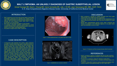
Case Description/Methods: A 56-year-old male presented with anemia. Initial endoscopic evaluation revealed several large gastric subepithelial lesions in the antrum and body of the stomach that were ulcerated. After biopsies showed only chronic inflammation, the patient was referred to our center for further evaluation with EUS. There were multiple bulky confluent submucosal hypoechoic lesions seen with ulcerated mucosa overlying involving the gastric body, angularis and proximal antrum. Given the large size it was difficult to determine from which wall layer they originated. Also, there were multiple malignant appearing lymph nodes seen adjacent to the stomach. Biopsies through both fine needle biopsy and forceps biopsies, the pathology report revealed low grade B-cell lymphoma with clonal plasmacytic differentiation, suggestive of marginal zone of mucosa-associated lymphoid tissue lymphoma (MALT).
Discussion: MALT lymphoma is typically an indolent low-grade tumor that usually presents with a more subtle endoscopic appearance. In this case of MALT lymphoma, the tumor was bulky, ulcerated and had metastasized to lymph nodes around the stomach. Also, there was no evidence of h. pylori infection. MALT lymphoma should considered on the differential diagnosis of any gastric lesion given the presentation can be so varied.

Disclosures:
Amirali Tavangar, MD1, David Cheung, MD2, Vamsi Vemireddy, MD1, Julie J. Oh, MD1, Jason Samarasena, MD, MBA, FACG3. A0701 - MALT Lymphoma: An Unlikely Diagnosis of Gastric Subepithelial Lesion, ACG 2022 Annual Scientific Meeting Abstracts. Charlotte, NC: American College of Gastroenterology.
1University of California Irvine, Orange, CA; 2UCI Medical Center, Orange, CA; 3UC Irvine, Orange, CA
Introduction: MALT lymphoma is one of the most common types of extra-nodal non-Hodgkin lymphomas which originate from marginal zone B-cells in the gastrointestinal mucosal-associated lymphoid tissues. Although the most frequent location which MALT can be found is stomach, it is a rare disorder. Most cases are associated with h. pylori infection of the stomach. It is typically a low-grade neoplasia, characterized by a dense lymphoid infiltration that invades and destroys gastric glands and results in the so-called “lymphoepithelial lesion”. We present a case that was an atypical presentation of Malt lymphoma.
Case Description/Methods: A 56-year-old male presented with anemia. Initial endoscopic evaluation revealed several large gastric subepithelial lesions in the antrum and body of the stomach that were ulcerated. After biopsies showed only chronic inflammation, the patient was referred to our center for further evaluation with EUS. There were multiple bulky confluent submucosal hypoechoic lesions seen with ulcerated mucosa overlying involving the gastric body, angularis and proximal antrum. Given the large size it was difficult to determine from which wall layer they originated. Also, there were multiple malignant appearing lymph nodes seen adjacent to the stomach. Biopsies through both fine needle biopsy and forceps biopsies, the pathology report revealed low grade B-cell lymphoma with clonal plasmacytic differentiation, suggestive of marginal zone of mucosa-associated lymphoid tissue lymphoma (MALT).
Discussion: MALT lymphoma is typically an indolent low-grade tumor that usually presents with a more subtle endoscopic appearance. In this case of MALT lymphoma, the tumor was bulky, ulcerated and had metastasized to lymph nodes around the stomach. Also, there was no evidence of h. pylori infection. MALT lymphoma should considered on the differential diagnosis of any gastric lesion given the presentation can be so varied.

Figure: EGD and EUS showing the lesion
Disclosures:
Amirali Tavangar indicated no relevant financial relationships.
David Cheung indicated no relevant financial relationships.
Vamsi Vemireddy indicated no relevant financial relationships.
Julie Oh indicated no relevant financial relationships.
Jason Samarasena: Conmed – Consultant. Docbot – Stock Options. Mauna Kea – Consultant. Olympus – Consultant. Ovesco – Consultant. Steris – Consultant.
Amirali Tavangar, MD1, David Cheung, MD2, Vamsi Vemireddy, MD1, Julie J. Oh, MD1, Jason Samarasena, MD, MBA, FACG3. A0701 - MALT Lymphoma: An Unlikely Diagnosis of Gastric Subepithelial Lesion, ACG 2022 Annual Scientific Meeting Abstracts. Charlotte, NC: American College of Gastroenterology.
