Back
Poster Session E - Tuesday Afternoon
E0714 - The Master of Disguise: Isolated Gastric Variceal Hemorrhage as a Complication of Endoscopic Gastric Biopsy
Tuesday, October 25, 2022
3:00 PM – 5:00 PM ET
Location: Crown Ballroom

Randy Leibowitz, DO
Mount Sinai St. Luke's and Mount Sinai Roosevelt
New York, NY
Presenting Author(s)
Randy Leibowitz, DO1, Frederick Rozenshteyn, MD2, Edward Lung, MD3, Abdallah Beano, MD4
1Mount Sinai St. Luke's and Mount Sinai Roosevelt, New York, NY; 2Icahn School of Medicine at Mount Sinai Morningside-West, New York, NY; 3Mount Sinai Morningside and Mount Sinai West, New York, NY; 4Mount Sinai Beth Israel, Morningside and West, New York, NY
Introduction: Splenic vein thrombosis (SVT) causes left-sided portal hypertension that can result in the formation of gastric varices. Gastric varices can be isolated or contiguous with esophageal varices. Isolated gastric varices may be difficult to distinguish from gastric rugae. They are responsible for 10-30% of all variceal bleeds.
Case Description/Methods: A 61-year old male with a past medical history of coronary artery disease on aspirin and clopidogrel presented to the emergency department with 2 days of black tarry stools and associated dyspnea and palpitations. 3 weeks ago, he saw an outpatient gastroenterologist to evaluate new onset anemia (hemoglobin 10 mg/dL) and an unexplained 25-pound weight loss over 6 months. He underwent esophagogastroduodenoscopy (EGD) 2 days prior to presentation in which a protruding lesion in the gastric fundus was biopsied.
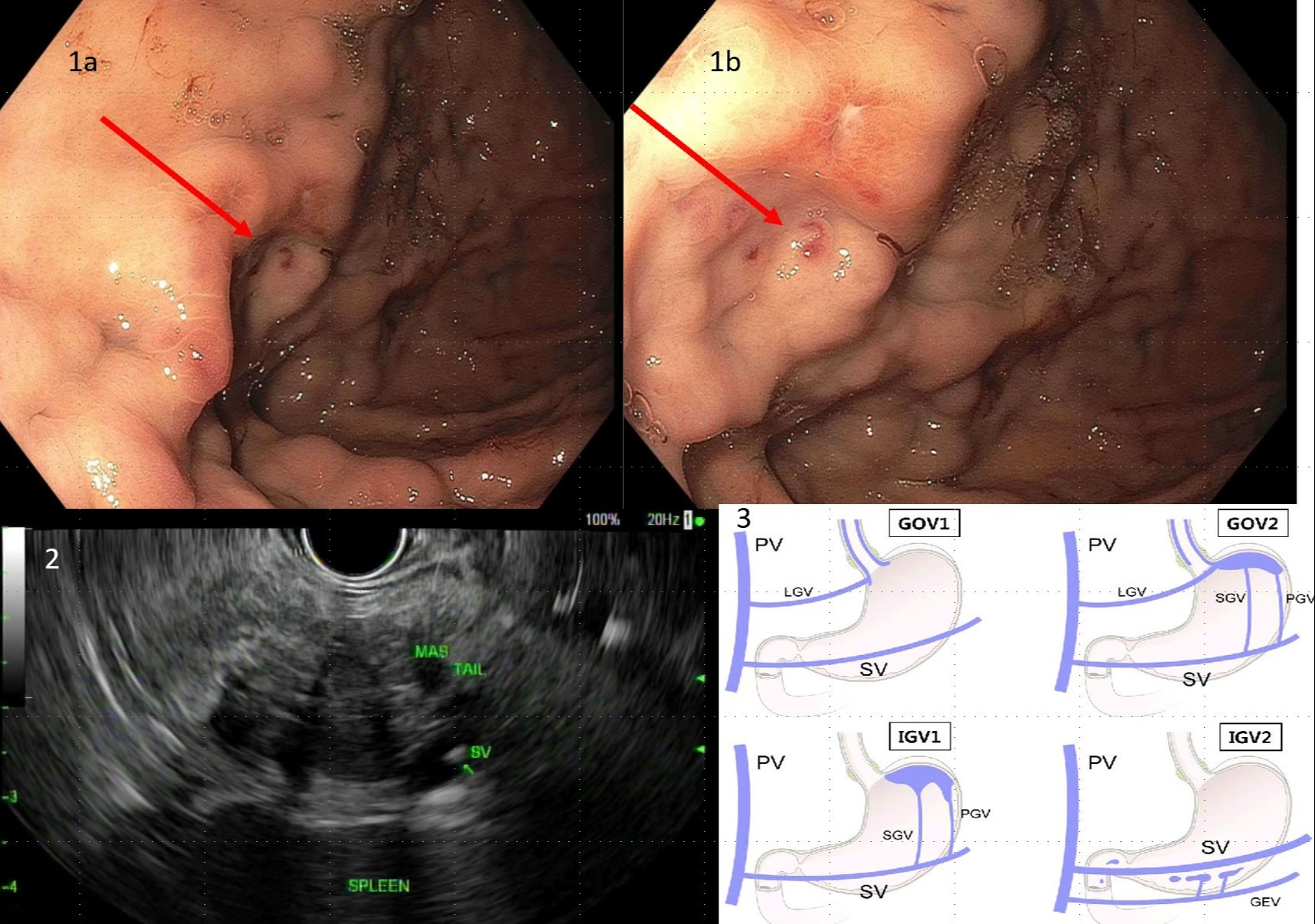
On initial presentation, he was hemodynamically stable with epigastric tenderness on physical exam. Initially, his hemoglobin was 8.1 mg/dL, which further decreased to 6.6 mg/dL that evening requiring transfusion. Computed tomography of the abdomen with IV contrast showed a 4.6cm pancreatic tail lesion with associated splenic vein thrombosis. The patient underwent EGD with endoscopic ultrasound (EUS) which revealed a fundal isolated gastric varix with stigmata of recent bleeding. EUS guided biopsy results of the pancreatic tail lesion demonstrated a well-differentiated neuroendocrine tumor.
Discussion: Gastric varices (GV) are categorized based on their location. Gastroesophageal varices (GOV) include GOV1, extending from the esophagus to the lesser curvature of the stomach, and GOV2, extending from the esophagus to the greater curvature. Isolated gastric varices (IGV) include IGV1 in the fundus, while IGV2 are located at ectopic gastric sites including the antrum, body, pylorus, incisura and duodenum. The most common gastric varix is GOV1 (74%). IGV1 comprises 8% of total GV and 78% of IGV. A prospective cohort study by Sarin et al. noted IGV1 to bleed at a much higher rate (78%) than IGV2 (9%). Gastric vein obliteration is the most successful modality in treating acute bleeds and harbors the lowest rebleeding rate (22%).
Gastric varices have a similar bleeding risk to esophageal varices (25%) and are more difficult to treat. For patients with risk factors for SVT (pancreatitis, cirrhosis and other prothrombotic states) who present with a GI bleed, IGV should be included in the differential.

Disclosures:
Randy Leibowitz, DO1, Frederick Rozenshteyn, MD2, Edward Lung, MD3, Abdallah Beano, MD4. E0714 - The Master of Disguise: Isolated Gastric Variceal Hemorrhage as a Complication of Endoscopic Gastric Biopsy, ACG 2022 Annual Scientific Meeting Abstracts. Charlotte, NC: American College of Gastroenterology.
1Mount Sinai St. Luke's and Mount Sinai Roosevelt, New York, NY; 2Icahn School of Medicine at Mount Sinai Morningside-West, New York, NY; 3Mount Sinai Morningside and Mount Sinai West, New York, NY; 4Mount Sinai Beth Israel, Morningside and West, New York, NY
Introduction: Splenic vein thrombosis (SVT) causes left-sided portal hypertension that can result in the formation of gastric varices. Gastric varices can be isolated or contiguous with esophageal varices. Isolated gastric varices may be difficult to distinguish from gastric rugae. They are responsible for 10-30% of all variceal bleeds.
Case Description/Methods: A 61-year old male with a past medical history of coronary artery disease on aspirin and clopidogrel presented to the emergency department with 2 days of black tarry stools and associated dyspnea and palpitations. 3 weeks ago, he saw an outpatient gastroenterologist to evaluate new onset anemia (hemoglobin 10 mg/dL) and an unexplained 25-pound weight loss over 6 months. He underwent esophagogastroduodenoscopy (EGD) 2 days prior to presentation in which a protruding lesion in the gastric fundus was biopsied.
On initial presentation, he was hemodynamically stable with epigastric tenderness on physical exam. Initially, his hemoglobin was 8.1 mg/dL, which further decreased to 6.6 mg/dL that evening requiring transfusion. Computed tomography of the abdomen with IV contrast showed a 4.6cm pancreatic tail lesion with associated splenic vein thrombosis. The patient underwent EGD with endoscopic ultrasound (EUS) which revealed a fundal isolated gastric varix with stigmata of recent bleeding. EUS guided biopsy results of the pancreatic tail lesion demonstrated a well-differentiated neuroendocrine tumor.
Discussion: Gastric varices (GV) are categorized based on their location. Gastroesophageal varices (GOV) include GOV1, extending from the esophagus to the lesser curvature of the stomach, and GOV2, extending from the esophagus to the greater curvature. Isolated gastric varices (IGV) include IGV1 in the fundus, while IGV2 are located at ectopic gastric sites including the antrum, body, pylorus, incisura and duodenum. The most common gastric varix is GOV1 (74%). IGV1 comprises 8% of total GV and 78% of IGV. A prospective cohort study by Sarin et al. noted IGV1 to bleed at a much higher rate (78%) than IGV2 (9%). Gastric vein obliteration is the most successful modality in treating acute bleeds and harbors the lowest rebleeding rate (22%).
Gastric varices have a similar bleeding risk to esophageal varices (25%) and are more difficult to treat. For patients with risk factors for SVT (pancreatitis, cirrhosis and other prothrombotic states) who present with a GI bleed, IGV should be included in the differential.

Figure: Figure 1a,b: Endoscopic image of isolated gastric varix with stigmata of recent bleeding; Figure 2: EUS image of the pancreatic tail mass and the splenic vein with visible thrombosis; Figure 3: Anatomy of gastroesophageal varices and Isolated gastric varices
SV=Splenic vein, PV=Portal vein, LGV=Left gastric vein, GEV=Gastroepiploic vein, SGV= Short gastric vein, PGV=Posterior gastric vein
SV=Splenic vein, PV=Portal vein, LGV=Left gastric vein, GEV=Gastroepiploic vein, SGV= Short gastric vein, PGV=Posterior gastric vein
Disclosures:
Randy Leibowitz indicated no relevant financial relationships.
Frederick Rozenshteyn indicated no relevant financial relationships.
Edward Lung indicated no relevant financial relationships.
Abdallah Beano indicated no relevant financial relationships.
Randy Leibowitz, DO1, Frederick Rozenshteyn, MD2, Edward Lung, MD3, Abdallah Beano, MD4. E0714 - The Master of Disguise: Isolated Gastric Variceal Hemorrhage as a Complication of Endoscopic Gastric Biopsy, ACG 2022 Annual Scientific Meeting Abstracts. Charlotte, NC: American College of Gastroenterology.
