Back
Poster Session E - Tuesday Afternoon
E0728 - Unusual Cause of Bleeding From an Underlying Glomus Tumor
Tuesday, October 25, 2022
3:00 PM – 5:00 PM ET
Location: Crown Ballroom

Ria Kundu, MD
Banner Health
Mesa, AZ
Presenting Author(s)
Aida Rezaie, MD1, Ria Kundu, MD2, Kayvon Sotoudeh, MD1, Brett Hughes, MD1, Hadiatou Barry, MD, MPH1, Indu Srinivasan, MD3, Keng-Yu Chuang, MD3
1Creighton University, Phoenix, AZ; 2Banner Health, Mesa, AZ; 3Valleywise Health, Phoenix, AZ
Introduction: Glomus tumors (GTs) are rare, mostly benign mesenchymal neoplasm accounting for nearly 1% of all gastrointestinal soft tissue tumors. Commonly seen in distal extremities, they do rarely occur in the gastrointestinal tract where it most commonly involves the stomach, especially the antrum. These tumors lack specific symptoms and endoscopic findings making it hard to differentiate from other gastrointestinal submucosal tumors without resection. We present a rare case of gastric glomus tumor (GGT) in the antrum diagnosed in a patient presenting with melena.
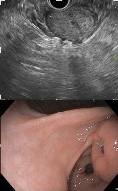
Case Description/Methods: 47-year- old male patient presented with melena. Upper endoscopy revealed a non-bleeding gastric ulcer with a visible vessel. Almost 3 cm submucosal lesion was seen at the site of the ulcer concerning for possible hematoma. Given the size of the lesion a follow up CT scan was performed 2 months later which showed persistent 3 cm submucosal circumscribed hyper enhancing lesion at the pylorus. The patient was then scheduled for endoscopic ultrasound which showed a hypoechoic lesion with discrete borders arising from the muscularis propria measuring 22 x 24 mm in maximal dimension. Fine needle biopsy of the lesion was performed using 22-gauge needle. Pathology revealed uniform round cells intimately associated with gaping vessels. The cells were reactive for vimentin and smooth muscle actin consistent with the diagnosis of glomus tumor. The patient was referred to surgery for excision.
Discussion: Glomus tumor of the stomach arise from the intramuscular layer and often present as a solitary submucosal lesion. GGTs usually present with epigastric discomfort, hematemesis, melena, nausea or vomiting or rarely as an incidental finding. Endoscopic ultrasound typically shows hypoechoic circumscribed lesions mostly arising from the fourth layer of the stomach. However biopsies are rarely conclusive thus making preoperative diagnosis challenging. Given its rare malignant potential, wide local surgical excision is the treatment of choice.

Disclosures:
Aida Rezaie, MD1, Ria Kundu, MD2, Kayvon Sotoudeh, MD1, Brett Hughes, MD1, Hadiatou Barry, MD, MPH1, Indu Srinivasan, MD3, Keng-Yu Chuang, MD3. E0728 - Unusual Cause of Bleeding From an Underlying Glomus Tumor, ACG 2022 Annual Scientific Meeting Abstracts. Charlotte, NC: American College of Gastroenterology.
1Creighton University, Phoenix, AZ; 2Banner Health, Mesa, AZ; 3Valleywise Health, Phoenix, AZ
Introduction: Glomus tumors (GTs) are rare, mostly benign mesenchymal neoplasm accounting for nearly 1% of all gastrointestinal soft tissue tumors. Commonly seen in distal extremities, they do rarely occur in the gastrointestinal tract where it most commonly involves the stomach, especially the antrum. These tumors lack specific symptoms and endoscopic findings making it hard to differentiate from other gastrointestinal submucosal tumors without resection. We present a rare case of gastric glomus tumor (GGT) in the antrum diagnosed in a patient presenting with melena.
Case Description/Methods: 47-year- old male patient presented with melena. Upper endoscopy revealed a non-bleeding gastric ulcer with a visible vessel. Almost 3 cm submucosal lesion was seen at the site of the ulcer concerning for possible hematoma. Given the size of the lesion a follow up CT scan was performed 2 months later which showed persistent 3 cm submucosal circumscribed hyper enhancing lesion at the pylorus. The patient was then scheduled for endoscopic ultrasound which showed a hypoechoic lesion with discrete borders arising from the muscularis propria measuring 22 x 24 mm in maximal dimension. Fine needle biopsy of the lesion was performed using 22-gauge needle. Pathology revealed uniform round cells intimately associated with gaping vessels. The cells were reactive for vimentin and smooth muscle actin consistent with the diagnosis of glomus tumor. The patient was referred to surgery for excision.
Discussion: Glomus tumor of the stomach arise from the intramuscular layer and often present as a solitary submucosal lesion. GGTs usually present with epigastric discomfort, hematemesis, melena, nausea or vomiting or rarely as an incidental finding. Endoscopic ultrasound typically shows hypoechoic circumscribed lesions mostly arising from the fourth layer of the stomach. However biopsies are rarely conclusive thus making preoperative diagnosis challenging. Given its rare malignant potential, wide local surgical excision is the treatment of choice.

Figure: Figure 1: Endoscopic ultrasound of a glomus tumor in the gastric antrum. Figure 2: Upper endoscopy of a glomus tumor with a central ulcer and visible vessel.
Disclosures:
Aida Rezaie indicated no relevant financial relationships.
Ria Kundu indicated no relevant financial relationships.
Kayvon Sotoudeh indicated no relevant financial relationships.
Brett Hughes indicated no relevant financial relationships.
Hadiatou Barry indicated no relevant financial relationships.
Indu Srinivasan indicated no relevant financial relationships.
Keng-Yu Chuang indicated no relevant financial relationships.
Aida Rezaie, MD1, Ria Kundu, MD2, Kayvon Sotoudeh, MD1, Brett Hughes, MD1, Hadiatou Barry, MD, MPH1, Indu Srinivasan, MD3, Keng-Yu Chuang, MD3. E0728 - Unusual Cause of Bleeding From an Underlying Glomus Tumor, ACG 2022 Annual Scientific Meeting Abstracts. Charlotte, NC: American College of Gastroenterology.
