Back
Poster Session D - Tuesday Morning
D0718 - A Rare Case of Glomus Tumor of the Stomach With Metastatic Spread to the Liver and Lungs
Tuesday, October 25, 2022
10:00 AM – 12:00 PM ET
Location: Crown Ballroom

Anirudh R. Damughatla, DO
Wayne State University/Detroit Medical Center
Detroit, MI
Presenting Author(s)
Anirudh R. Damughatla, DO1, Sai Vaishnavi Kamatham, MD1, Jasdeep S. Bathla, DO2, Mohamad I. Itani, MD3, Sarvani Surapaneni, MD1, Vanessa Milan-Ortiz, MD2, Anthony F. Shields, MD, PhD4
1Wayne State University/Detroit Medical Center, Detroit, MI; 2Detroit Medical Center/Wayne State University, Detroit, MI; 3Wayne State University / Detroit Medical Center, Detroit, MI; 4Karmanos Cancer Institute, Detroit, MI
Introduction: Glomus tumors are rare mesenchymal neoplasms arising from glomus bodies and occur mostly as benign tumors located in the subungual region of the digits. Glomus tumors of the gastrointestinal tract are rare, where there may be few or no glomus bodies, and the majority are benign, rarely demonstrating malignant behavior. We present a case of a metastatic glomus tumor of the stomach, which was initially deemed benign.
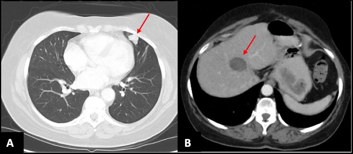
Case Description/Methods: We present a 72-year-old woman who underwent excision of a gastric mass with a partial gastrectomy in 2010, suspecting a Gastrointestinal stromal tumor (GIST). However, pathology findings were consistent with glomus tumor of uncertain malignant potential given borderline tumor size (2 cm) and increased mitotic activity ( >=5/50 HPF). She was not started on chemotherapy and was on surveillance. She was asymptomatic until 2016, when she presented with acute abdominal pain. Imaging showed multiple new necrotic metastases throughout the liver with pathology consistent with metastatic glomus tumor. She underwent radioembolization of the right hepatic and left hepatic artery in 2017 and continued to be symptom-free until 2019 when she presented with another episode of abdominal pain; imaging showed an increase in the number of liver lesions and underwent radioembolization with a good response. Repeat surveillance imaging in early 2020 showed a nodule in the lingula (shown in figure 1a). A CT-guided lung biopsy confirmed the diagnosis of an epithelioid neoplasm, consistent with a metastatic glomus tumor. Caris genomic testing did not show any apparent mutations, and no clear molecular targets for treatments were present. She had low ERCC1 and TOPO1, suggesting that oxaliplatin and irinotecan may be considered for treatment in case of disease progression.
Discussion: Our case highlights the importance of considering glomus tumors as part of the differentials in GI mesenchymal tumors despite GIST having the highest incidence. It is known that many GI mesenchymal tumors have visual and radiological similarities, and hence a biopsy is required. Although a majority of glomus tumors are benign, obtaining histological and immunohistochemical features is still imperative to make a definitive diagnosis to detect malignant glomus tumors with metastatic potential to visceral organs as they have a poor prognosis.

Disclosures:
Anirudh R. Damughatla, DO1, Sai Vaishnavi Kamatham, MD1, Jasdeep S. Bathla, DO2, Mohamad I. Itani, MD3, Sarvani Surapaneni, MD1, Vanessa Milan-Ortiz, MD2, Anthony F. Shields, MD, PhD4. D0718 - A Rare Case of Glomus Tumor of the Stomach With Metastatic Spread to the Liver and Lungs, ACG 2022 Annual Scientific Meeting Abstracts. Charlotte, NC: American College of Gastroenterology.
1Wayne State University/Detroit Medical Center, Detroit, MI; 2Detroit Medical Center/Wayne State University, Detroit, MI; 3Wayne State University / Detroit Medical Center, Detroit, MI; 4Karmanos Cancer Institute, Detroit, MI
Introduction: Glomus tumors are rare mesenchymal neoplasms arising from glomus bodies and occur mostly as benign tumors located in the subungual region of the digits. Glomus tumors of the gastrointestinal tract are rare, where there may be few or no glomus bodies, and the majority are benign, rarely demonstrating malignant behavior. We present a case of a metastatic glomus tumor of the stomach, which was initially deemed benign.
Case Description/Methods: We present a 72-year-old woman who underwent excision of a gastric mass with a partial gastrectomy in 2010, suspecting a Gastrointestinal stromal tumor (GIST). However, pathology findings were consistent with glomus tumor of uncertain malignant potential given borderline tumor size (2 cm) and increased mitotic activity ( >=5/50 HPF). She was not started on chemotherapy and was on surveillance. She was asymptomatic until 2016, when she presented with acute abdominal pain. Imaging showed multiple new necrotic metastases throughout the liver with pathology consistent with metastatic glomus tumor. She underwent radioembolization of the right hepatic and left hepatic artery in 2017 and continued to be symptom-free until 2019 when she presented with another episode of abdominal pain; imaging showed an increase in the number of liver lesions and underwent radioembolization with a good response. Repeat surveillance imaging in early 2020 showed a nodule in the lingula (shown in figure 1a). A CT-guided lung biopsy confirmed the diagnosis of an epithelioid neoplasm, consistent with a metastatic glomus tumor. Caris genomic testing did not show any apparent mutations, and no clear molecular targets for treatments were present. She had low ERCC1 and TOPO1, suggesting that oxaliplatin and irinotecan may be considered for treatment in case of disease progression.
Discussion: Our case highlights the importance of considering glomus tumors as part of the differentials in GI mesenchymal tumors despite GIST having the highest incidence. It is known that many GI mesenchymal tumors have visual and radiological similarities, and hence a biopsy is required. Although a majority of glomus tumors are benign, obtaining histological and immunohistochemical features is still imperative to make a definitive diagnosis to detect malignant glomus tumors with metastatic potential to visceral organs as they have a poor prognosis.

Figure: Figure 1: CT Thorax (1a) and abdomen (1b) w/contrast showing epithelioid neoplasm of the lingula and hypo-dense liver mass respectively.
Disclosures:
Anirudh Damughatla indicated no relevant financial relationships.
Sai Vaishnavi Kamatham indicated no relevant financial relationships.
Jasdeep Bathla indicated no relevant financial relationships.
Mohamad I. Itani indicated no relevant financial relationships.
Sarvani Surapaneni indicated no relevant financial relationships.
Vanessa Milan-Ortiz indicated no relevant financial relationships.
Anthony Shields: Alkermes – Grant/Research Support. Astellas – Grant/Research Support. Astra Zeneca – Grant/Research Support. Bayer – Grant/Research Support. Boehringer Ingelheim – Grant/Research Support. Boston Biomedical – Grant/Research Support. Caris Life Sciences – Advisor or Review Panel Member, Grant/Research Support, Speakers Bureau. Cogent Biosciences – Consultant. Daiichi Sankyo – Grant/Research Support. Eisai – Grant/Research Support. Esperas Pharma – Grant/Research Support. Exelexis – Grant/Research Support. Five Prime Therapeutics – Grant/Research Support. Gritstone bio – Grant/Research Support. H3 Biomedicine – Grant/Research Support. Hutchinson China MediTech – Grant/Research Support. Imaginab – Grant/Research Support. Incyte – Grant/Research Support. Jiangsu Alphamed – Grant/Research Support. LSK BioPartners – Grant/Research Support. Nouscom – Grant/Research Support. Nuvation Bio – Grant/Research Support. Repertoire Immune Medicines – Grant/Research Support. Seattle Genetics – Grant/Research Support. Shanghai Haihe – Grant/Research Support. SQZ Biotechnologies – Grant/Research Support. Telix Pharmaceuticals – Grant/Research Support. TopAlliance – Grant/Research Support. Xencor – Grant/Research Support.
Anirudh R. Damughatla, DO1, Sai Vaishnavi Kamatham, MD1, Jasdeep S. Bathla, DO2, Mohamad I. Itani, MD3, Sarvani Surapaneni, MD1, Vanessa Milan-Ortiz, MD2, Anthony F. Shields, MD, PhD4. D0718 - A Rare Case of Glomus Tumor of the Stomach With Metastatic Spread to the Liver and Lungs, ACG 2022 Annual Scientific Meeting Abstracts. Charlotte, NC: American College of Gastroenterology.
