Back
Poster Session C - Monday Afternoon
C0701 - An Atypical Presentation of Burkitt’s Lymphoma
Monday, October 24, 2022
3:00 PM – 5:00 PM ET
Location: Crown Ballroom

Lyam Ciccone, MD
NYU Langone Medical Center/ NYCHH-Woodhull Medical Center
Brooklyn, NY
Presenting Author(s)
Lyam Ciccone, MD1, Yvette Achuo-Egbe, MD, MPH, MS2, Ashley Maranino, MD2
1NYU Langone Medical Center/ NYCHH-Woodhull Medical Center, Brooklyn, NY; 2New York Medical College/ NYCHH- Metropolitan Hospital Center, New York, NY
Introduction: Burkitt’s lymphoma is an aggressive non-Hodgkin lymphoma, uncommon in adults and has 3 subtypes. Immunodeficiency-associated subtype is primarily linked to HIV infection accounting for 30%-50% of HIV associated lymphoma. Abdominal involvement can be found in this subtype. We present a case of an atypical presentation of Burkitt’s lymphoma.
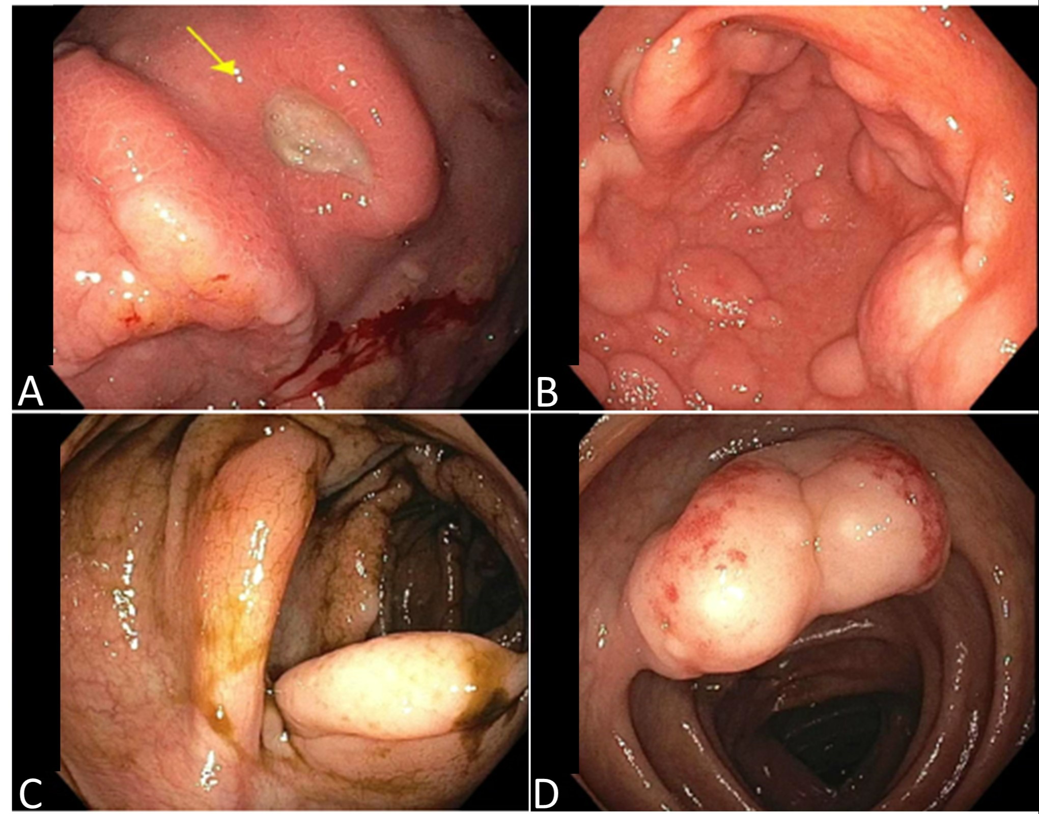
Case Description/Methods: A 38-year-old man with no significant past medical history who presented to the emergency room with nausea, vomiting, bloating, non-bloody diarrhea, unintentional weight loss of 15 lbs, fevers, chills, diaphoresis and decreased appetite for 1 month. On physical examination, there was evidence of abdominal distension with ascites and hepatomegaly, and bilateral lower extremity edema. Laboratory tests showed elevated AST of 67 U/L, ALT of 42 U/L, lactic acid of 3.7 mmol/L and LDH of 1,091 U/L. CT showed extensive periaortic, pericaval common and external iliac chain lymphadenopathy; circumferential small bowel wall thickening and associated contrast enhancement suggesting inflammation. Further workup revealed positivity for HIV sub-type B. Patient underwent EGD and colonoscopy. On EGD, a non-bleeding cratered ulcer was seen in the lesser curvature of the stomach and numerous, large, confluent papules noted on the entire stomach, bulb and second portion of the duodenum. On colonoscopy, a 7 mm lipomatous-appearing submucosal nodule was found in the ileocecal valve, a 30 mm polypoid non-obstructing mass in the proximal transverse colon and a 20 mm polypoid lesion in the proximal transverse colon. All lesions were biopsied. (Figure-1 A,B,C,D). Biopsy results revealed composite High-grade B-Cell Lymphoma (HBCL) with MYC+ Burkitt lymphoma.
Discussion: Burkitt’s lymphoma is a relatively rare malignancy in adults accounting for about 1-5% of all non-Hodgkin lymphomas in this population. Mesenteric and retroperitoneal lymph node involvement is common. Small bowel involvement may be seen on imaging as circumferential thickening, aneurysmal dilatation or as an intraluminal polyp/mass. Based on our patient’s vague initial presentation, multiple differentials were considered including gastrointestinal lymphoma, neuroendocrine tumors, inflammatory bowel disease or infectious disease. A robust differential was only achieved after the finding of periaortic and pericaval lymphadenopathy which directed our focus towards lymphatic malignancy and the later discovered presence of HIV infection further supported the suspicion of Burkitt’s lymphoma.

Disclosures:
Lyam Ciccone, MD1, Yvette Achuo-Egbe, MD, MPH, MS2, Ashley Maranino, MD2. C0701 - An Atypical Presentation of Burkitt’s Lymphoma, ACG 2022 Annual Scientific Meeting Abstracts. Charlotte, NC: American College of Gastroenterology.
1NYU Langone Medical Center/ NYCHH-Woodhull Medical Center, Brooklyn, NY; 2New York Medical College/ NYCHH- Metropolitan Hospital Center, New York, NY
Introduction: Burkitt’s lymphoma is an aggressive non-Hodgkin lymphoma, uncommon in adults and has 3 subtypes. Immunodeficiency-associated subtype is primarily linked to HIV infection accounting for 30%-50% of HIV associated lymphoma. Abdominal involvement can be found in this subtype. We present a case of an atypical presentation of Burkitt’s lymphoma.
Case Description/Methods: A 38-year-old man with no significant past medical history who presented to the emergency room with nausea, vomiting, bloating, non-bloody diarrhea, unintentional weight loss of 15 lbs, fevers, chills, diaphoresis and decreased appetite for 1 month. On physical examination, there was evidence of abdominal distension with ascites and hepatomegaly, and bilateral lower extremity edema. Laboratory tests showed elevated AST of 67 U/L, ALT of 42 U/L, lactic acid of 3.7 mmol/L and LDH of 1,091 U/L. CT showed extensive periaortic, pericaval common and external iliac chain lymphadenopathy; circumferential small bowel wall thickening and associated contrast enhancement suggesting inflammation. Further workup revealed positivity for HIV sub-type B. Patient underwent EGD and colonoscopy. On EGD, a non-bleeding cratered ulcer was seen in the lesser curvature of the stomach and numerous, large, confluent papules noted on the entire stomach, bulb and second portion of the duodenum. On colonoscopy, a 7 mm lipomatous-appearing submucosal nodule was found in the ileocecal valve, a 30 mm polypoid non-obstructing mass in the proximal transverse colon and a 20 mm polypoid lesion in the proximal transverse colon. All lesions were biopsied. (Figure-1 A,B,C,D). Biopsy results revealed composite High-grade B-Cell Lymphoma (HBCL) with MYC+ Burkitt lymphoma.
Discussion: Burkitt’s lymphoma is a relatively rare malignancy in adults accounting for about 1-5% of all non-Hodgkin lymphomas in this population. Mesenteric and retroperitoneal lymph node involvement is common. Small bowel involvement may be seen on imaging as circumferential thickening, aneurysmal dilatation or as an intraluminal polyp/mass. Based on our patient’s vague initial presentation, multiple differentials were considered including gastrointestinal lymphoma, neuroendocrine tumors, inflammatory bowel disease or infectious disease. A robust differential was only achieved after the finding of periaortic and pericaval lymphadenopathy which directed our focus towards lymphatic malignancy and the later discovered presence of HIV infection further supported the suspicion of Burkitt’s lymphoma.

Figure: Figure-1. Endoscopy and Colonoscopy findings. A – Gastric ulcer in body of the stomach. B – Papules and nodules and pre-pyloric stomach. C – Polypoid lesion in transverse colon. D – Mass in transverse colon.
Disclosures:
Lyam Ciccone indicated no relevant financial relationships.
Yvette Achuo-Egbe indicated no relevant financial relationships.
Ashley Maranino indicated no relevant financial relationships.
Lyam Ciccone, MD1, Yvette Achuo-Egbe, MD, MPH, MS2, Ashley Maranino, MD2. C0701 - An Atypical Presentation of Burkitt’s Lymphoma, ACG 2022 Annual Scientific Meeting Abstracts. Charlotte, NC: American College of Gastroenterology.
