Back
Poster Session C - Monday Afternoon
C0700 - Gastric Cancer Presenting With Bilateral Ureteral Obstruction
Monday, October 24, 2022
3:00 PM – 5:00 PM ET
Location: Crown Ballroom

Khaled Elfert, MD, MRCP
SBH Health System
New York, NY
Presenting Author(s)
Khaled Elfert, MD, MRCP1, Bashar Tanous, MD2, Xheni Deda, MD3, Ali Rahil, MD2, Azizullah A. Beran, MD4
1SBH Health System, New York, NY; 2Hamad Medical Corporation, Doha, Ad Dawhah, Qatar; 3SBH Health System, Bronx, NY; 4The University of Toledo, Toledo, OH
Introduction: Ureteral obstruction can occur due to metastases in patients with pelvic malignancy. However, its occurrence in non-pelvic malignancies, e.g., gastric cancer, is very rare and has a poor prognosis. Here, we report a patient who initially presented with bilateral ureteral obstruction and was subsequently diagnosed with gastric cancer.
Case Description/Methods: A 58-year-old female patient with a history of diabetes mellitus and hypertension presented to the hospital due to bilateral flank pain and anuria; she also reported early satiety and non-intentional weight loss of 10 kgs. Her physical examination was remarkable for bilateral flank tenderness.
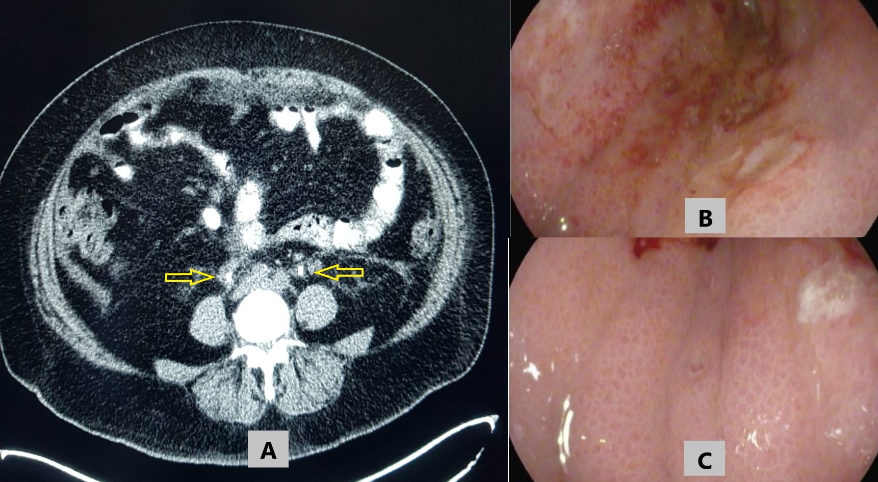
Blood tests showed creatinine of 656.96 umol/L and urea of 16.9 mmol/L. Her baseline creatinine was normal. Renal ultrasound revealed bilateral hydronephrosis. CT of the abdomen with contrast was notable for bilateral hydronephrosis with no renal or ureteric stones, and diffuse thickening of the stomach wall with omental nodules and ascites. She underwent bilateral double J-stent placement (Figure 1a), and subsequently, her kidney function improved.
EGD (Figure 1b,c) showed edematous and thickened gastric mucosal folds that didn't flatten with insufflation. Gastric histopathological examination reported poorly differentiated adenocarcinoma (MMR intact, and negative for HER2/neu and H. pylori). Furthermore, PET scan revealed metastatic lymph nodes above and below the diaphragm with peritoneal involvement. The patient was started on FOLFOX chemotherapy regimen and Nivolumab. A follow-up abdominal CT scan after she received five cycles of chemotherapy showed moderate tumor regression.
Discussion: Here, we described a patient with gastric cancer that presented with bilateral ureteral obstruction. Different mechanisms for ureteral obstruction in gastric cancer have been described. One of the mechanisms is the direct extension of the primary tumor, peritoneal deposits or lymph nodes to the ureters. Other mechanisms include retroperitoneal fibrosis as a reaction to the malignant cells and distant metastases to the ureter from the primary gastric tumor. In our patient, the most likely etiology of the ureteral obstruction is an extension of the peritoneal metastases to the ureter. Our case provides an example of an unusual clinical presentation of gastric cancer and adds to the growing literature about acute bilateral obstructive uropathy as an initial presentation of gastric cancer. Physicians should be aware of such a rare presentation of gastric cancer.

Disclosures:
Khaled Elfert, MD, MRCP1, Bashar Tanous, MD2, Xheni Deda, MD3, Ali Rahil, MD2, Azizullah A. Beran, MD4. C0700 - Gastric Cancer Presenting With Bilateral Ureteral Obstruction, ACG 2022 Annual Scientific Meeting Abstracts. Charlotte, NC: American College of Gastroenterology.
1SBH Health System, New York, NY; 2Hamad Medical Corporation, Doha, Ad Dawhah, Qatar; 3SBH Health System, Bronx, NY; 4The University of Toledo, Toledo, OH
Introduction: Ureteral obstruction can occur due to metastases in patients with pelvic malignancy. However, its occurrence in non-pelvic malignancies, e.g., gastric cancer, is very rare and has a poor prognosis. Here, we report a patient who initially presented with bilateral ureteral obstruction and was subsequently diagnosed with gastric cancer.
Case Description/Methods: A 58-year-old female patient with a history of diabetes mellitus and hypertension presented to the hospital due to bilateral flank pain and anuria; she also reported early satiety and non-intentional weight loss of 10 kgs. Her physical examination was remarkable for bilateral flank tenderness.
Blood tests showed creatinine of 656.96 umol/L and urea of 16.9 mmol/L. Her baseline creatinine was normal. Renal ultrasound revealed bilateral hydronephrosis. CT of the abdomen with contrast was notable for bilateral hydronephrosis with no renal or ureteric stones, and diffuse thickening of the stomach wall with omental nodules and ascites. She underwent bilateral double J-stent placement (Figure 1a), and subsequently, her kidney function improved.
EGD (Figure 1b,c) showed edematous and thickened gastric mucosal folds that didn't flatten with insufflation. Gastric histopathological examination reported poorly differentiated adenocarcinoma (MMR intact, and negative for HER2/neu and H. pylori). Furthermore, PET scan revealed metastatic lymph nodes above and below the diaphragm with peritoneal involvement. The patient was started on FOLFOX chemotherapy regimen and Nivolumab. A follow-up abdominal CT scan after she received five cycles of chemotherapy showed moderate tumor regression.
Discussion: Here, we described a patient with gastric cancer that presented with bilateral ureteral obstruction. Different mechanisms for ureteral obstruction in gastric cancer have been described. One of the mechanisms is the direct extension of the primary tumor, peritoneal deposits or lymph nodes to the ureters. Other mechanisms include retroperitoneal fibrosis as a reaction to the malignant cells and distant metastases to the ureter from the primary gastric tumor. In our patient, the most likely etiology of the ureteral obstruction is an extension of the peritoneal metastases to the ureter. Our case provides an example of an unusual clinical presentation of gastric cancer and adds to the growing literature about acute bilateral obstructive uropathy as an initial presentation of gastric cancer. Physicians should be aware of such a rare presentation of gastric cancer.

Figure: A) Computed Tomography scan image demonstrating the thickened ureters (yellow arrows) mainly on the right side with the double J-stents seen inside their lumen. Endoscopic images of the gastric body (B, C) demonstrate thickened, edematous gastric mucosal folds, with a waffle-like appearance, that did not flatten with insufflation consistent with linitis plastica.
Disclosures:
Khaled Elfert indicated no relevant financial relationships.
Bashar Tanous indicated no relevant financial relationships.
Xheni Deda indicated no relevant financial relationships.
Azizullah Beran indicated no relevant financial relationships.
Khaled Elfert, MD, MRCP1, Bashar Tanous, MD2, Xheni Deda, MD3, Ali Rahil, MD2, Azizullah A. Beran, MD4. C0700 - Gastric Cancer Presenting With Bilateral Ureteral Obstruction, ACG 2022 Annual Scientific Meeting Abstracts. Charlotte, NC: American College of Gastroenterology.
