Back
Poster Session E - Tuesday Afternoon
E0662 - Laparoscopic-Assisted Enteroscopy with “Shar Pei” Technique for Resection of Deep Small Bowel Polyps
Tuesday, October 25, 2022
3:00 PM – 5:00 PM ET
Location: Crown Ballroom

Muaaz Masood, MD
Medical College of Georgia at Augusta University
Augusta, GA
Presenting Author(s)
Award: Presidential Poster Award
Muaaz Masood, MD1, Aaron Bolduc, MD2, John Erikson L. Yap, MD3
1Medical College of Georgia at Augusta University, Augusta, GA; 2Augusta University, Augusta, GA; 3Augusta University Medical College of Georiga, Augusta, GA
Introduction: Balloon enteroscopy (BE) has transformed the management of small bowel disease. However, BE still has limits in terms of reaching deeper parts of the small bowel. Laparoscopic-assisted enteroscopy (LAE) has emerged as an effective procedure for small bowel polyps. We present 2 cases of LAE using a novel Shar Pei technique.
Case Description/Methods: Case 1: A 71-year-old female who underwent bidirectional endoscopy and video capsule endoscopy (VCE) for work-up of iron deficiency anemia. VCE revealed a non-obstructing polypoid lesion with minimal oozing in the proximal small bowel at 15% small bowel transit time (SBTT). Anterograde BE revealed a flat, 10-mm polyp in the proximal jejunum. Due to unstable positioning, the polyp was removed incompletely via piecemeal cold snare polypectomy (CSP). She subsequently underwent LAE with Shar Pei technique to pleat the small bowel over the enteroscope until the polyp was reached. The polyp was removed en bloc via endoscopic mucosal resection (EMR).
Case 2: A 23-year-old female with Peutz-Jeghers syndrome was found to have 2 polypoid lesions in the small bowel on surveillance imaging. VCE revealed 2 polyps at 32% and 82% SBTT, respectively. The polyps were not reachable via anterograde and retrograde BE. A LAE was performed and the small bowel was pleated using the Shar Pei technique until a 50 mm semi-pedunculated polyp was visualized by the endoscope 320 cm from the Ligament of Treitz. The polyp was removed en bloc via EMR. Next, a colonoscope was advanced to the terminal ileum which was pleated laparoscopically until three polyps were seen 25 cm past the ileocecal valve that were removed with en bloc EMR.
Discussion: LAE is an effective, minimally invasive technique for the management of deep small bowel pathology. LAE preserves the mucosal integrity eliminating the need for anastomoses. LAE has a high detection rate for small intestinal disease and is generally safe. No immediate complications were noted in our cases. Our novel Shar Pei technique was named after the dog breed whose characteristic wrinkled skin resembles the folds of the small bowel. The technique entailed laparoscopically advancing the small bowel over the endoscope while it remained stationary. The proximal end of the small bowel was stabilized during polyp resection to secure the endoscope and prevent telescoping backwards. LAE with the Shar Pei technique is a novel, promising tool for the diagnosis and treatment of small bowel polyps.

Disclosures:
Muaaz Masood, MD1, Aaron Bolduc, MD2, John Erikson L. Yap, MD3. E0662 - Laparoscopic-Assisted Enteroscopy with “Shar Pei” Technique for Resection of Deep Small Bowel Polyps, ACG 2022 Annual Scientific Meeting Abstracts. Charlotte, NC: American College of Gastroenterology.
Muaaz Masood, MD1, Aaron Bolduc, MD2, John Erikson L. Yap, MD3
1Medical College of Georgia at Augusta University, Augusta, GA; 2Augusta University, Augusta, GA; 3Augusta University Medical College of Georiga, Augusta, GA
Introduction: Balloon enteroscopy (BE) has transformed the management of small bowel disease. However, BE still has limits in terms of reaching deeper parts of the small bowel. Laparoscopic-assisted enteroscopy (LAE) has emerged as an effective procedure for small bowel polyps. We present 2 cases of LAE using a novel Shar Pei technique.
Case Description/Methods: Case 1: A 71-year-old female who underwent bidirectional endoscopy and video capsule endoscopy (VCE) for work-up of iron deficiency anemia. VCE revealed a non-obstructing polypoid lesion with minimal oozing in the proximal small bowel at 15% small bowel transit time (SBTT). Anterograde BE revealed a flat, 10-mm polyp in the proximal jejunum. Due to unstable positioning, the polyp was removed incompletely via piecemeal cold snare polypectomy (CSP). She subsequently underwent LAE with Shar Pei technique to pleat the small bowel over the enteroscope until the polyp was reached. The polyp was removed en bloc via endoscopic mucosal resection (EMR).
Case 2: A 23-year-old female with Peutz-Jeghers syndrome was found to have 2 polypoid lesions in the small bowel on surveillance imaging. VCE revealed 2 polyps at 32% and 82% SBTT, respectively. The polyps were not reachable via anterograde and retrograde BE. A LAE was performed and the small bowel was pleated using the Shar Pei technique until a 50 mm semi-pedunculated polyp was visualized by the endoscope 320 cm from the Ligament of Treitz. The polyp was removed en bloc via EMR. Next, a colonoscope was advanced to the terminal ileum which was pleated laparoscopically until three polyps were seen 25 cm past the ileocecal valve that were removed with en bloc EMR.
Discussion: LAE is an effective, minimally invasive technique for the management of deep small bowel pathology. LAE preserves the mucosal integrity eliminating the need for anastomoses. LAE has a high detection rate for small intestinal disease and is generally safe. No immediate complications were noted in our cases. Our novel Shar Pei technique was named after the dog breed whose characteristic wrinkled skin resembles the folds of the small bowel. The technique entailed laparoscopically advancing the small bowel over the endoscope while it remained stationary. The proximal end of the small bowel was stabilized during polyp resection to secure the endoscope and prevent telescoping backwards. LAE with the Shar Pei technique is a novel, promising tool for the diagnosis and treatment of small bowel polyps.

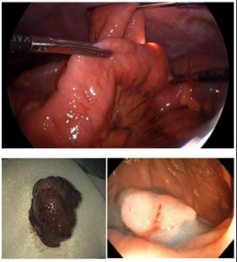
Figure: Panel 1. (Top) Laparoscopic-assisted enteroscopy with Shar Pei technique was utilized to telescope the small bowel over the endoscope until the 50-mm polyp was successfully reached at 320 cm from the Ligament of Treitz and resected with EMR (bottom left). This technique was also used to resect a 10-mm jejunal polyp with en bloc EMR (bottom right).
Disclosures:
Muaaz Masood indicated no relevant financial relationships.
Aaron Bolduc indicated no relevant financial relationships.
John Erikson Yap indicated no relevant financial relationships.
Muaaz Masood, MD1, Aaron Bolduc, MD2, John Erikson L. Yap, MD3. E0662 - Laparoscopic-Assisted Enteroscopy with “Shar Pei” Technique for Resection of Deep Small Bowel Polyps, ACG 2022 Annual Scientific Meeting Abstracts. Charlotte, NC: American College of Gastroenterology.

