Back
Poster Session D - Tuesday Morning
D0663 - Primary T-Cell Lymphoma of the Small Intestine Presenting as Perforation Peritonitis
Tuesday, October 25, 2022
10:00 AM – 12:00 PM ET
Location: Crown Ballroom

Razan Aljaras, MD
Indiana University School of Medicine
Indianapolis, IN
Presenting Author(s)
Razan Aljaras, MD1, Astin Worden, MD1, Eleazar E. Montalvan-Sanchez, MD1, Renato Beas, MD2, Ahmad Karkash, MD1, Rawan Aljaras, MD1
1Indiana University School of Medicine, Indianapolis, IN; 2Indiana University, Indianapolis, IN
Introduction: Intestinal T-cell lymphomas are rare primary T-cell lymphomas. The two most common types are enteropathy-associated T-cell lymphoma (EATL) and monomorphic epitheliotropic intestinal T-cell lymphoma (MEITL). MEITL was newly defined by the 2016 revision of the World Health Organization, it was previously known as EATL type II. EATL is associated with celiac disease whereas MEITL is not. Both types of lymphoma usually present with non-specific symptoms.
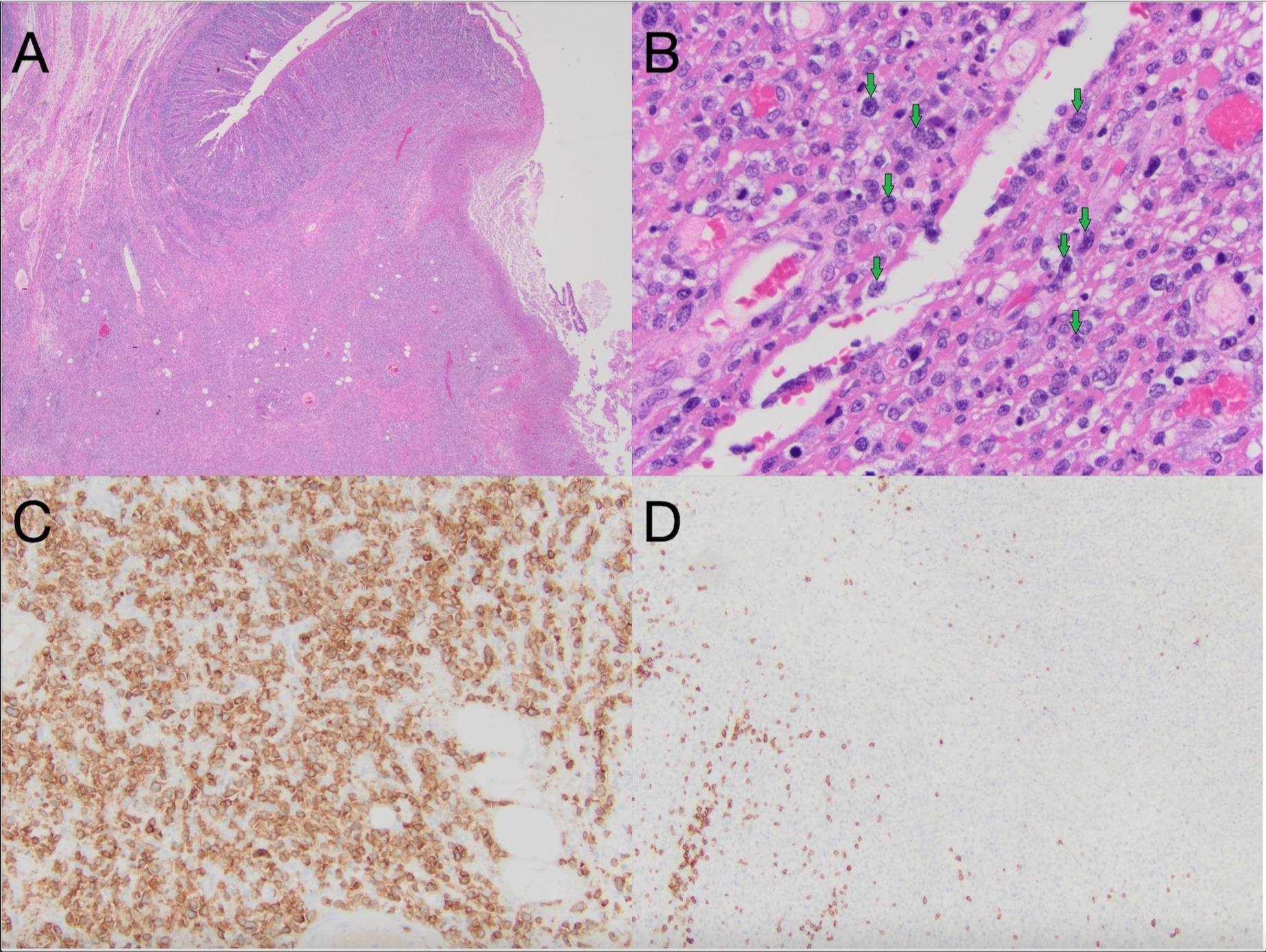
Case Description/Methods: We present the case of a 65 year old female patient with no known relevant medical history presented with an acute onset of abdominal pain. X-ray showed pneumoperitoneum and the patient was taken to surgery. During surgery, a perforation in the small bowel was identified and a segment of the small bowel was resected. Histologic examination of the specimen showed full-thickness involvement by atypical medium-sized lymphoid cells with areas of necrosis. The overlying epithelium showed increased intraepithelial lymphocytes. Immunostains showed that the neoplastic cells are positive for CD2, CD3, and CD8 with aberrant loss of CD5 and CD7. The findings were most consistent with monomorphic epitheliotropic intestinal T-cell lymphoma. Histologic images with select immunostains are seen in the attached figure.
Discussion: We present a rare case of intestinal T-cell lymphoma that was interestingly first diagnosed after it perforated the small bowel. MEITL carries an incidence of 0.25% of all malignant lymphomas and <5% of all digestive tract malignant lymphomas. It is unfortunately a very aggressive malignancy that carries a poor prognosis and a high mortality rate. Oftentimes, MEITL also represents a diagnostic challenge. Careful histologic examination is key in diagnosing this entity of disease.

Disclosures:
Razan Aljaras, MD1, Astin Worden, MD1, Eleazar E. Montalvan-Sanchez, MD1, Renato Beas, MD2, Ahmad Karkash, MD1, Rawan Aljaras, MD1. D0663 - Primary T-Cell Lymphoma of the Small Intestine Presenting as Perforation Peritonitis, ACG 2022 Annual Scientific Meeting Abstracts. Charlotte, NC: American College of Gastroenterology.
1Indiana University School of Medicine, Indianapolis, IN; 2Indiana University, Indianapolis, IN
Introduction: Intestinal T-cell lymphomas are rare primary T-cell lymphomas. The two most common types are enteropathy-associated T-cell lymphoma (EATL) and monomorphic epitheliotropic intestinal T-cell lymphoma (MEITL). MEITL was newly defined by the 2016 revision of the World Health Organization, it was previously known as EATL type II. EATL is associated with celiac disease whereas MEITL is not. Both types of lymphoma usually present with non-specific symptoms.
Case Description/Methods: We present the case of a 65 year old female patient with no known relevant medical history presented with an acute onset of abdominal pain. X-ray showed pneumoperitoneum and the patient was taken to surgery. During surgery, a perforation in the small bowel was identified and a segment of the small bowel was resected. Histologic examination of the specimen showed full-thickness involvement by atypical medium-sized lymphoid cells with areas of necrosis. The overlying epithelium showed increased intraepithelial lymphocytes. Immunostains showed that the neoplastic cells are positive for CD2, CD3, and CD8 with aberrant loss of CD5 and CD7. The findings were most consistent with monomorphic epitheliotropic intestinal T-cell lymphoma. Histologic images with select immunostains are seen in the attached figure.
Discussion: We present a rare case of intestinal T-cell lymphoma that was interestingly first diagnosed after it perforated the small bowel. MEITL carries an incidence of 0.25% of all malignant lymphomas and <5% of all digestive tract malignant lymphomas. It is unfortunately a very aggressive malignancy that carries a poor prognosis and a high mortality rate. Oftentimes, MEITL also represents a diagnostic challenge. Careful histologic examination is key in diagnosing this entity of disease.

Figure: A- A transmural lymphoid infiltrate is seen with mucosal ulceration (20x).
B- The tumor cells are medium-sized to large (400x).
C- The tumor cells are positive for CD3.
D- The tumor cells show aberrant loss of CD5.
B- The tumor cells are medium-sized to large (400x).
C- The tumor cells are positive for CD3.
D- The tumor cells show aberrant loss of CD5.
Disclosures:
Razan Aljaras indicated no relevant financial relationships.
Astin Worden indicated no relevant financial relationships.
Eleazar Montalvan-Sanchez indicated no relevant financial relationships.
Renato Beas indicated no relevant financial relationships.
Ahmad Karkash indicated no relevant financial relationships.
Rawan Aljaras indicated no relevant financial relationships.
Razan Aljaras, MD1, Astin Worden, MD1, Eleazar E. Montalvan-Sanchez, MD1, Renato Beas, MD2, Ahmad Karkash, MD1, Rawan Aljaras, MD1. D0663 - Primary T-Cell Lymphoma of the Small Intestine Presenting as Perforation Peritonitis, ACG 2022 Annual Scientific Meeting Abstracts. Charlotte, NC: American College of Gastroenterology.
