Back
Poster Session E - Tuesday Afternoon
E0535 - Hepatic Cerebrospinal Fluid Pseudocyst: A Rare Complication of Ventriculoperitoneal Shunt
Tuesday, October 25, 2022
3:00 PM – 5:00 PM ET
Location: Crown Ballroom

Muhammad Nadeem Yousaf, MD
University of Missouri
Columbia, MO
Presenting Author(s)
Muhammad Nadeem Yousaf, MD1, Haider Abbas Naqvi, MD2, Shriya Kane, 3, Fizah Chaudhary, MD1, Jason Hawksworth, MD4, Vikram Nayar, MD4, Thomas W. Faust, MD, MBE5
1University of Missouri, Columbia, MO; 2MedStar Health, Baltimore, MD; 3Georgetown University School of Medicine, Washington, DC; 4MedStar Georgetown University Hospital, Washington, DC; 5James D. Eason Transplant Institute, Methodist University, University of Tennessee Health Science Center, Memphis, TN
Introduction: Ventriculoperitoneal shunts (VPS) are commonly used in the management of hydrocephalus to drain cerebrospinal fluid (CSF) into the peritoneal cavity. The placement of VPS has prolonged the survival of patients with hydrocephalus. Abdominal pseudocysts are long-term complication of VPS that are identified later in life. Hepatic CSF pseudocyst is a rare long-term complication of VPS that should be differentiated from other cystic lesions of liver.
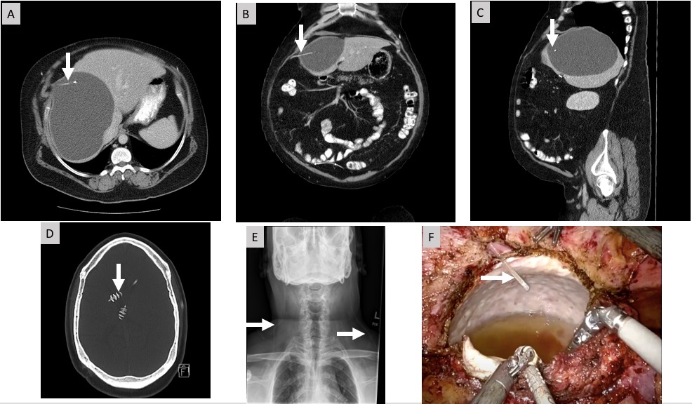
Case Description/Methods: A 49-year-old man with intellectual disability from congenital hydrocephalus s/p placement of right and left-sided VPS at age of 3 months and 7 years respectively presented with exertional dyspnea and abdominal distension. On presentation his vitals were unremarkable except tachycardic (115/min)., He had abdominal distention, hepatomegaly but no tenderness. Initial labs showed D-dimer 2.20 mcg/mL, AST 27 u/L, ALT 38 u/L, alkaline phosphate 126 u/L and total bilirubin 0.5 mg/dL. Chest CT with IV contrast was negative for pulmonary embolism, however revealed a large 18x13x13.5 cm cyst in right hepatic lobe. CT abdomen and pelvis demonstrated a 17.5x12.6x12.7 cm cystic lesion in the right hepatic lobe with the tip of VP shunt catheter within cyst cavity. Hepatobiliary nuclear scan was unremarkable for any biliary leak or sphincter of oddi dysfunction. CT head was negative for any acute abnormalities. Shunt series x-rays were negative for disruption of VPS catheter. Robotic laparoscopic cyst fenestration with partial hepatectomy was performed and catheter was repositioned to the right lower quadrant of abdomen. Patient was discharged home two days later with significant reduction of cyst size on follow up imaging.
Discussion: This case illustrates a rare complication of VPS that result in hepatic CSF pseudocyst. If hydrocephalus is absent, patients with hepatic CSF pseudocyst are asymptomatic at earlier stages., or they may present with abdominal pain, distention, or palpable right upper quadrant abdominal mass. A subset of patients with hepatic CSF pseudocyst are complicated with bacterial or parasitic infection and present with abdominal pain, distention, or right upper quadrant mass. Abdominal ultrasound and CT scan assist in diagnosis by identifying the tip of VPS catheter in the pseudocyst cavity. Asymptomatic patients are managed conservatively while surgical repositioning of VPS catheter with or without cyst fenestration or surgical excision of cyst may be required for complete resolution of symptoms.

Disclosures:
Muhammad Nadeem Yousaf, MD1, Haider Abbas Naqvi, MD2, Shriya Kane, 3, Fizah Chaudhary, MD1, Jason Hawksworth, MD4, Vikram Nayar, MD4, Thomas W. Faust, MD, MBE5. E0535 - Hepatic Cerebrospinal Fluid Pseudocyst: A Rare Complication of Ventriculoperitoneal Shunt, ACG 2022 Annual Scientific Meeting Abstracts. Charlotte, NC: American College of Gastroenterology.
1University of Missouri, Columbia, MO; 2MedStar Health, Baltimore, MD; 3Georgetown University School of Medicine, Washington, DC; 4MedStar Georgetown University Hospital, Washington, DC; 5James D. Eason Transplant Institute, Methodist University, University of Tennessee Health Science Center, Memphis, TN
Introduction: Ventriculoperitoneal shunts (VPS) are commonly used in the management of hydrocephalus to drain cerebrospinal fluid (CSF) into the peritoneal cavity. The placement of VPS has prolonged the survival of patients with hydrocephalus. Abdominal pseudocysts are long-term complication of VPS that are identified later in life. Hepatic CSF pseudocyst is a rare long-term complication of VPS that should be differentiated from other cystic lesions of liver.
Case Description/Methods: A 49-year-old man with intellectual disability from congenital hydrocephalus s/p placement of right and left-sided VPS at age of 3 months and 7 years respectively presented with exertional dyspnea and abdominal distension. On presentation his vitals were unremarkable except tachycardic (115/min)., He had abdominal distention, hepatomegaly but no tenderness. Initial labs showed D-dimer 2.20 mcg/mL, AST 27 u/L, ALT 38 u/L, alkaline phosphate 126 u/L and total bilirubin 0.5 mg/dL. Chest CT with IV contrast was negative for pulmonary embolism, however revealed a large 18x13x13.5 cm cyst in right hepatic lobe. CT abdomen and pelvis demonstrated a 17.5x12.6x12.7 cm cystic lesion in the right hepatic lobe with the tip of VP shunt catheter within cyst cavity. Hepatobiliary nuclear scan was unremarkable for any biliary leak or sphincter of oddi dysfunction. CT head was negative for any acute abnormalities. Shunt series x-rays were negative for disruption of VPS catheter. Robotic laparoscopic cyst fenestration with partial hepatectomy was performed and catheter was repositioned to the right lower quadrant of abdomen. Patient was discharged home two days later with significant reduction of cyst size on follow up imaging.
Discussion: This case illustrates a rare complication of VPS that result in hepatic CSF pseudocyst. If hydrocephalus is absent, patients with hepatic CSF pseudocyst are asymptomatic at earlier stages., or they may present with abdominal pain, distention, or palpable right upper quadrant abdominal mass. A subset of patients with hepatic CSF pseudocyst are complicated with bacterial or parasitic infection and present with abdominal pain, distention, or right upper quadrant mass. Abdominal ultrasound and CT scan assist in diagnosis by identifying the tip of VPS catheter in the pseudocyst cavity. Asymptomatic patients are managed conservatively while surgical repositioning of VPS catheter with or without cyst fenestration or surgical excision of cyst may be required for complete resolution of symptoms.

Figure: Figure 1: CT of abdomen showing a large right hepatic lobe cyst with evidence of tip of right VPS catheter within the cavity of the cyst (arrows) on transverse (A), axial (B) and lateral (C) views. CT scan of the head (D) showing VPS catheters both in right and left lateral ventricles without evidence of ventricular dilation. Shunt series x-ray (E) shows now distortion of VPS catheter. Robotic laparoscopic cyst fenestration (F) shows VPS catheter within cyst cavity containing CSF.
Disclosures:
Muhammad Nadeem Yousaf indicated no relevant financial relationships.
Haider Abbas Naqvi indicated no relevant financial relationships.
Shriya Kane indicated no relevant financial relationships.
Fizah Chaudhary indicated no relevant financial relationships.
Jason Hawksworth indicated no relevant financial relationships.
Vikram Nayar indicated no relevant financial relationships.
Thomas Faust indicated no relevant financial relationships.
Muhammad Nadeem Yousaf, MD1, Haider Abbas Naqvi, MD2, Shriya Kane, 3, Fizah Chaudhary, MD1, Jason Hawksworth, MD4, Vikram Nayar, MD4, Thomas W. Faust, MD, MBE5. E0535 - Hepatic Cerebrospinal Fluid Pseudocyst: A Rare Complication of Ventriculoperitoneal Shunt, ACG 2022 Annual Scientific Meeting Abstracts. Charlotte, NC: American College of Gastroenterology.
