Back
Poster Session E - Tuesday Afternoon
E0456 - A Rare Case of Small Cell Lung Cancer in the Posterior Mediastinum Diagnosed With Endoscopic Ultrasound-Guided Fine Needle Biopsy
Tuesday, October 25, 2022
3:00 PM – 5:00 PM ET
Location: Crown Ballroom

Jamil M. Shah, MD
The Brooklyn Hospital Center
Brooklyn, NY
Presenting Author(s)
Jamil M. Shah, MD, Pratyusha Tirumanisetty, MD, Suut Gokturk, MD, Jasparit Minhas, MD, Philip Xiao, MD, Madhavi Reddy, MD, Ardeshir Hakami-Kermani, MD, Derrick Cheung, MD
The Brooklyn Hospital Center, Brooklyn, NY
Introduction: Lung cancer is the leading cause of cancer mortality worldwide, accounting for 2 million diagnoses and 1.8 million deaths in 2020.1 10-15% of all lung cancers are small cell lung cancer (SCLC), an aggressive form that grows more rapidly and metastasizes more easily than NSCLC. Thus, in patients with suspected lung cancer, a tissue diagnosis is crucial. Here, we present a unique case of a man with a posterior mediastinal mass for whom obtaining a tissue diagnosis presented a diagnostic challenge.
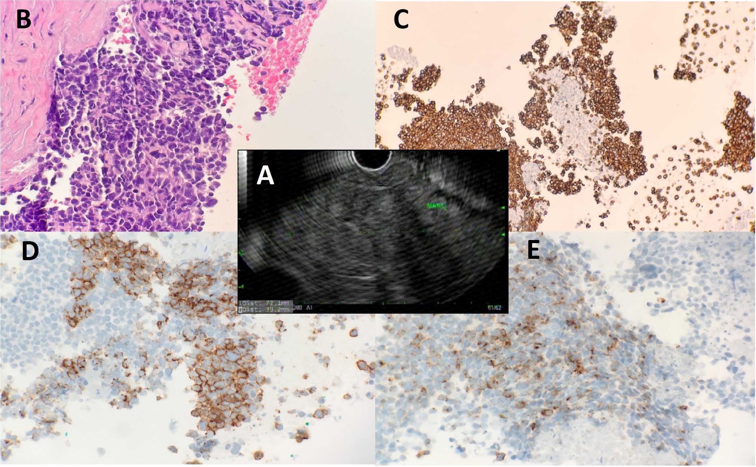
Case Description/Methods: A 49 y/o M, with a 30 pack-year smoking history, presented with worsening left-sided chest pain and a 10-pound unintentional weight loss over 2 months. Vital signs and physical exam were unremarkable. Labs demonstrated a mild anemia (Hb 11-12, MCV 82) and elevated inflammatory markers (ESR 54, CRP 70.9). CT scan demonstrated a 12.0x10.7x8.4 cm hypodense soft tissue density mass in the left posterior mediastinum, extending from the level of T1 to T7 vertebral bodies. The mass displaced the trachea, esophagus, and adjacent vessels. Several enlarged mediastinal lymph nodes were seen, the largest measuring up to 1.5 cm. However, there was no lymphadenopathy seen elsewhere and no convincing evidence of metastatic disease in the abdomen or pelvis. CT surgery recommended a CT-guided biopsy with IR vs. endoscopic bronchial US-guided biopsy. IR was unable to find a safe access point to go in for the biopsy as there was no window, as the lung was seen overlying the mass. The patient was discharged, and he returned the next week for outpatient EUS-FNB, which demonstrated a 72x39 mm mediastinal mass (Fig 1A). EUS-FNB was performed and EUS-guided transesophageal biopsies were performed. He was also found to have erosive gastritis, healing superficial sub-cm gastric ulcers, and multiple healing superficial duodenal ulcers. Pathology results demonstrated SCLC (Fig 1B-E) and H. pylori. He was treated with standard quadruple therapy, referred to his PCP to confirm eradication in 6 weeks, and referred to Oncology. For stage III-b SCLC treatment, he completed external beam RT and chemotherapy. Repeat PETCT demonstrated a decrease in the residual mass to 5.5x3.5x5.8 cm.
Discussion: For patients presenting with a posterior mediastinal mass, for whom IR may not be able to perform a biopsy, obtaining a tissue diagnosis may be a diagnostic challenge. EUS-FNB provides a useful tool to assess the mediastinum and can provide timely diagnosis and staging of patients with rare forms of lung cancer.

Disclosures:
Jamil M. Shah, MD, Pratyusha Tirumanisetty, MD, Suut Gokturk, MD, Jasparit Minhas, MD, Philip Xiao, MD, Madhavi Reddy, MD, Ardeshir Hakami-Kermani, MD, Derrick Cheung, MD. E0456 - A Rare Case of Small Cell Lung Cancer in the Posterior Mediastinum Diagnosed With Endoscopic Ultrasound-Guided Fine Needle Biopsy, ACG 2022 Annual Scientific Meeting Abstracts. Charlotte, NC: American College of Gastroenterology.
The Brooklyn Hospital Center, Brooklyn, NY
Introduction: Lung cancer is the leading cause of cancer mortality worldwide, accounting for 2 million diagnoses and 1.8 million deaths in 2020.1 10-15% of all lung cancers are small cell lung cancer (SCLC), an aggressive form that grows more rapidly and metastasizes more easily than NSCLC. Thus, in patients with suspected lung cancer, a tissue diagnosis is crucial. Here, we present a unique case of a man with a posterior mediastinal mass for whom obtaining a tissue diagnosis presented a diagnostic challenge.
Case Description/Methods: A 49 y/o M, with a 30 pack-year smoking history, presented with worsening left-sided chest pain and a 10-pound unintentional weight loss over 2 months. Vital signs and physical exam were unremarkable. Labs demonstrated a mild anemia (Hb 11-12, MCV 82) and elevated inflammatory markers (ESR 54, CRP 70.9). CT scan demonstrated a 12.0x10.7x8.4 cm hypodense soft tissue density mass in the left posterior mediastinum, extending from the level of T1 to T7 vertebral bodies. The mass displaced the trachea, esophagus, and adjacent vessels. Several enlarged mediastinal lymph nodes were seen, the largest measuring up to 1.5 cm. However, there was no lymphadenopathy seen elsewhere and no convincing evidence of metastatic disease in the abdomen or pelvis. CT surgery recommended a CT-guided biopsy with IR vs. endoscopic bronchial US-guided biopsy. IR was unable to find a safe access point to go in for the biopsy as there was no window, as the lung was seen overlying the mass. The patient was discharged, and he returned the next week for outpatient EUS-FNB, which demonstrated a 72x39 mm mediastinal mass (Fig 1A). EUS-FNB was performed and EUS-guided transesophageal biopsies were performed. He was also found to have erosive gastritis, healing superficial sub-cm gastric ulcers, and multiple healing superficial duodenal ulcers. Pathology results demonstrated SCLC (Fig 1B-E) and H. pylori. He was treated with standard quadruple therapy, referred to his PCP to confirm eradication in 6 weeks, and referred to Oncology. For stage III-b SCLC treatment, he completed external beam RT and chemotherapy. Repeat PETCT demonstrated a decrease in the residual mass to 5.5x3.5x5.8 cm.
Discussion: For patients presenting with a posterior mediastinal mass, for whom IR may not be able to perform a biopsy, obtaining a tissue diagnosis may be a diagnostic challenge. EUS-FNB provides a useful tool to assess the mediastinum and can provide timely diagnosis and staging of patients with rare forms of lung cancer.

Figure: A) A posterior mediastinal mass vs. esophageal mass measuring 72.1 mm by 39.2 mm was identified status-post EUS with FNB x 6. This mass appeared to arise from the muscularis propria of the esophagus; however, an extrinsic mediastinal mass could not be definitively ruled out due to the severe compressive effects of the mass on the esophagus.
Mediastinal mass, needle biopsy:
-Minute fragments of malignant neoplasm with necrosis.
-Tumor cells are positive for AE1/3, CAM5.2, Chromogranin, Synaptophysin, CD56 and negative for p40, CD3, CD20, and CD45. Ki-67 demonstrats about 60% positivity. Combined with morphological features, this immunoprofile supports the diagnosis of small cell carcinoma.
B) Hematoxylin and eosin (H&E) stain. C) CAM5.2. D) CD56. E) Chromogranin.
Disclosures:
Jamil Shah indicated no relevant financial relationships.
Pratyusha Tirumanisetty indicated no relevant financial relationships.
Suut Gokturk indicated no relevant financial relationships.
Jasparit Minhas indicated no relevant financial relationships.
Philip Xiao indicated no relevant financial relationships.
Madhavi Reddy indicated no relevant financial relationships.
Ardeshir Hakami-Kermani indicated no relevant financial relationships.
Derrick Cheung indicated no relevant financial relationships.
Jamil M. Shah, MD, Pratyusha Tirumanisetty, MD, Suut Gokturk, MD, Jasparit Minhas, MD, Philip Xiao, MD, Madhavi Reddy, MD, Ardeshir Hakami-Kermani, MD, Derrick Cheung, MD. E0456 - A Rare Case of Small Cell Lung Cancer in the Posterior Mediastinum Diagnosed With Endoscopic Ultrasound-Guided Fine Needle Biopsy, ACG 2022 Annual Scientific Meeting Abstracts. Charlotte, NC: American College of Gastroenterology.
