Back
Poster Session C - Monday Afternoon
C0465 - Disseminated Cryptococcosis Masquerading as Large Abdominal Mass Concerning for Pancreatic Malignancy in HIV-Positive Patient
Monday, October 24, 2022
3:00 PM – 5:00 PM ET
Location: Crown Ballroom

Monica Dzwonkowski, DO
Geisinger Medical Center
Danville, PA
Presenting Author(s)
Award: Presidential Poster Award
Monica Dzwonkowski, DO1, Umair Iqbal, MD1, Nihit Shah, MD2, Bradley D. Confer, DO1
1Geisinger Medical Center, Danville, PA; 2Geisinger Health System, Danville, PA
Introduction: Gastrointestinal cryptococcus is a rare occurrence in HIV-positive patients. Symptoms may be nonspecific thus the differential for abdominal masses must remain broad. We report a patient with HIV who presented with abdominal pain and watery diarrhea, found to have a large abdominal mass on computed tomography initially thought to be lymphoma or a primary pancreatic tumor. He underwent endoscopic ultrasound-guided (EUS) biopsy of the mass which was consistent with a cryptococcoma.
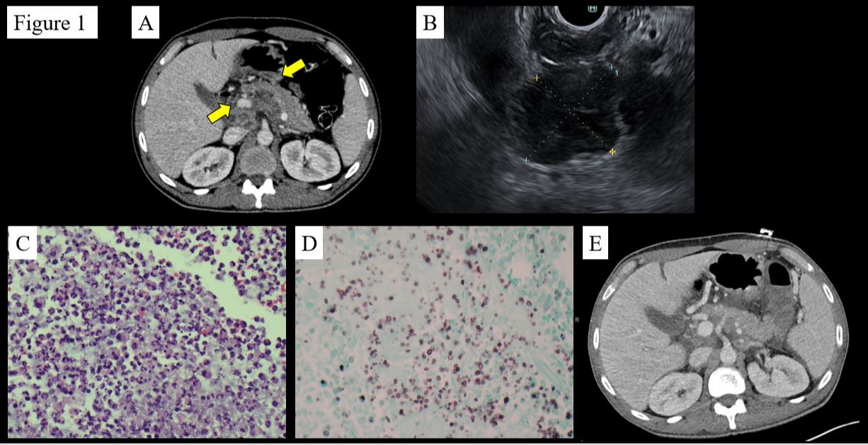
Case Description/Methods: A 55-year-old male presented with severe, diffuse abdominal pain for one week duration associated with profuse watery, non-bloody diarrhea and intermittent chills. Medical history included HIV for 30 years with poor compliance with antiretroviral regimen, recent pneumocystis pneumonia and previous cryptococcal meningitis. Physical exam revealed an ill appearing, cachectic male with abdominal tenderness. Labs revealed a white blood cell count of 18.5 K/uL (ref: 4.0-10.8 K/uL) with 84% neutrophils (ref: 40-75%), sodium of 132 mmol/L (ref: 135-146 mmol/L) and potassium 2.7 mmol/L (ref: 3.5-5.1 mmol/L). His viral load was 109,385 copies/mL (ref: neg copies/mL) and absolute CD4 count was 56 lymphocytes/uL (ref: 330-1520 lymphocytes/uL). Liver chemistries were unremarkable. Stool studies for regional pathogens, ova and parasites, and clostridium difficile were negative. A CT scan of the abdomen and pelvis revealed ill-defined cystic/necrotic upper abdominal lymphadenopathy with mass effect and possible invasion of the adjacent liver (Fig 1A). The pancreas and caudate lobe of the liver were inseparable from the mass which raised concern for primary pancreatic tumor or lymphoma. He underwent EUS-guided biopsy of the peripancreatic mass (Fig 1B) with pathology consistent with a cryptococcoma (Fig 1C, 1D). He was started on amphotericin B and transitioned to high dose fluconazole due to adverse effects. Repeat imaging revealed significant reduction of the mesenteric lymphadenopathy (Fig 1E). He was discharged in stable condition and remained on fluconazole indefinitely.
Discussion: Cryptococcal infection remains the second most common cause of acquired immunodeficiency syndrome related mortality after tuberculosis. The most common sites of cryptococcal infection are the lungs, brain, eyes, or central nervous system; abdominal dissemination is rare. Biopsy via EUS can be useful in diagnosis of cryptococcoma. Early identification and treatment of cryptococcal infections can increase survival rates.

Disclosures:
Monica Dzwonkowski, DO1, Umair Iqbal, MD1, Nihit Shah, MD2, Bradley D. Confer, DO1. C0465 - Disseminated Cryptococcosis Masquerading as Large Abdominal Mass Concerning for Pancreatic Malignancy in HIV-Positive Patient, ACG 2022 Annual Scientific Meeting Abstracts. Charlotte, NC: American College of Gastroenterology.
Monica Dzwonkowski, DO1, Umair Iqbal, MD1, Nihit Shah, MD2, Bradley D. Confer, DO1
1Geisinger Medical Center, Danville, PA; 2Geisinger Health System, Danville, PA
Introduction: Gastrointestinal cryptococcus is a rare occurrence in HIV-positive patients. Symptoms may be nonspecific thus the differential for abdominal masses must remain broad. We report a patient with HIV who presented with abdominal pain and watery diarrhea, found to have a large abdominal mass on computed tomography initially thought to be lymphoma or a primary pancreatic tumor. He underwent endoscopic ultrasound-guided (EUS) biopsy of the mass which was consistent with a cryptococcoma.
Case Description/Methods: A 55-year-old male presented with severe, diffuse abdominal pain for one week duration associated with profuse watery, non-bloody diarrhea and intermittent chills. Medical history included HIV for 30 years with poor compliance with antiretroviral regimen, recent pneumocystis pneumonia and previous cryptococcal meningitis. Physical exam revealed an ill appearing, cachectic male with abdominal tenderness. Labs revealed a white blood cell count of 18.5 K/uL (ref: 4.0-10.8 K/uL) with 84% neutrophils (ref: 40-75%), sodium of 132 mmol/L (ref: 135-146 mmol/L) and potassium 2.7 mmol/L (ref: 3.5-5.1 mmol/L). His viral load was 109,385 copies/mL (ref: neg copies/mL) and absolute CD4 count was 56 lymphocytes/uL (ref: 330-1520 lymphocytes/uL). Liver chemistries were unremarkable. Stool studies for regional pathogens, ova and parasites, and clostridium difficile were negative. A CT scan of the abdomen and pelvis revealed ill-defined cystic/necrotic upper abdominal lymphadenopathy with mass effect and possible invasion of the adjacent liver (Fig 1A). The pancreas and caudate lobe of the liver were inseparable from the mass which raised concern for primary pancreatic tumor or lymphoma. He underwent EUS-guided biopsy of the peripancreatic mass (Fig 1B) with pathology consistent with a cryptococcoma (Fig 1C, 1D). He was started on amphotericin B and transitioned to high dose fluconazole due to adverse effects. Repeat imaging revealed significant reduction of the mesenteric lymphadenopathy (Fig 1E). He was discharged in stable condition and remained on fluconazole indefinitely.
Discussion: Cryptococcal infection remains the second most common cause of acquired immunodeficiency syndrome related mortality after tuberculosis. The most common sites of cryptococcal infection are the lungs, brain, eyes, or central nervous system; abdominal dissemination is rare. Biopsy via EUS can be useful in diagnosis of cryptococcoma. Early identification and treatment of cryptococcal infections can increase survival rates.

Figure: Fig 1A: CT image showing ill-defined cystic/necrotic upper abdominal lymphadenopathy with
mass effect and possible invasion of the adjacent liver
Fig 1B: Endoscopic ultrasound imaging showing peripancreatic mass
Fig 1C: Hematoxylin and Eosin cellblock at 40X magnification showing abundant neutrophils and
necrotic debris
Fig 1D. Grocott’s Methenamine Silver stain showing fungal yeast with narrow base budding
Fig 1E. Significant reduction of the mesenteric lymphadenopathy after antifungal therapy
initiation
Disclosures:
Monica Dzwonkowski indicated no relevant financial relationships.
Umair Iqbal indicated no relevant financial relationships.
Nihit Shah indicated no relevant financial relationships.
Bradley Confer indicated no relevant financial relationships.
Monica Dzwonkowski, DO1, Umair Iqbal, MD1, Nihit Shah, MD2, Bradley D. Confer, DO1. C0465 - Disseminated Cryptococcosis Masquerading as Large Abdominal Mass Concerning for Pancreatic Malignancy in HIV-Positive Patient, ACG 2022 Annual Scientific Meeting Abstracts. Charlotte, NC: American College of Gastroenterology.

