Back
Poster Session C - Monday Afternoon
C0435 - Malignant Peritoneal Mesothelioma Seen on MRI Enterography During IBD Colitis Investigation in a 32-Year-Old Non-Asbestos Exposed Female Healthcare Worker
Monday, October 24, 2022
3:00 PM – 5:00 PM ET
Location: Crown Ballroom
- YS
Yecheskel Schneider, MD
St. Luke's University Health Network
Bethlehem, PA
Presenting Author(s)
Paul Dumitrescu, BS1, Matthew Record, BA1, Yecheskel Schneider, MD2
1Temple University School of Medicine, Bethlehem, PA; 2St. Luke's University Health Network, Bethlehem, PA
Introduction: This case describes an unexpected diagnosis of a malignant peritoneal mesothelioma (MPM) in a 32 y/o female physician without any known asbestos exposure. She presented to our department with a prior diagnosis of non-celiac gluten intolerance and intermittent but worsening left-sided abdominal pain for over 20 years. Several CTs suggested findings associated with IBD-related etiology and IBD specialist consultation was advised. After negative EGD/colonoscopy, an MRI Enterography revealed a small enhancing nodule suggestive of peritoneal malignancy. Pathology report after biopsy revealed a biphasic MPM. This case highlights the insidious nature of MPM and adds to the body of literature suggestive of IBD-mimicking symptomatology associated with this disease.
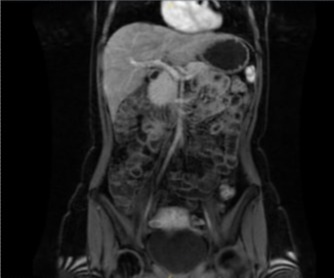
Case Description/Methods: A 32 y/o F presented with chronic intermittent left-sided abdominal pain and gluten intolerance. Throughout her life, she reports 2-week periods of abdominal pain that is usually dull and sometimes sharp. The pain is focused LUQ with occasional LLQ pain. This is worsened by acute angle bending and deep palpation to the area. The patient still experiences pain despite a gluten-free diet. EGD is negative for celiac disease. CT imaging showed a left-sided pericolonic abscess and wall thickening of the lower descending and proximal sigmoid colon. An ill-defined soft tissue density in the left paracolic gutter was also seen and classified as a reactive lymph node. Abscess underwent CT aspiration; follow-up CT noted a new focal area of segmental wall thickening in the ascending colon with an inflamed diverticulum. IBD consult was recommended where negative EGD/colonoscopy resulted in MRI Enterography referral and results suggestive of peritoneal malignancy (figure 1).
Pathologic examination confirmed the presence of MPM, biphasic type (epithelial/spindle) with cytologic atypia and invasive growth being consistent with malignancy. The patient is on nivolumab, ipilimumab, carboplatin, pemetrexed and leuprolide for the next 3 months and considering debulking.
Discussion: MPM presenting with decades long history of LUQ/LLQ pain exacerbations is rarely seen. CT results led to consideration of IBD etiology, as wall thickening, inflamed diverticulum, and a timeline suggestive of IBD was described. The lack of asbestos exposure highlighted the unlikely MPM differential. Prompt advanced imaging was key to diagnosis. Due to the insidious nature of MPM and poor outcomes, further exploration of timely advanced imaging is warranted.

Disclosures:
Paul Dumitrescu, BS1, Matthew Record, BA1, Yecheskel Schneider, MD2. C0435 - Malignant Peritoneal Mesothelioma Seen on MRI Enterography During IBD Colitis Investigation in a 32-Year-Old Non-Asbestos Exposed Female Healthcare Worker, ACG 2022 Annual Scientific Meeting Abstracts. Charlotte, NC: American College of Gastroenterology.
1Temple University School of Medicine, Bethlehem, PA; 2St. Luke's University Health Network, Bethlehem, PA
Introduction: This case describes an unexpected diagnosis of a malignant peritoneal mesothelioma (MPM) in a 32 y/o female physician without any known asbestos exposure. She presented to our department with a prior diagnosis of non-celiac gluten intolerance and intermittent but worsening left-sided abdominal pain for over 20 years. Several CTs suggested findings associated with IBD-related etiology and IBD specialist consultation was advised. After negative EGD/colonoscopy, an MRI Enterography revealed a small enhancing nodule suggestive of peritoneal malignancy. Pathology report after biopsy revealed a biphasic MPM. This case highlights the insidious nature of MPM and adds to the body of literature suggestive of IBD-mimicking symptomatology associated with this disease.
Case Description/Methods: A 32 y/o F presented with chronic intermittent left-sided abdominal pain and gluten intolerance. Throughout her life, she reports 2-week periods of abdominal pain that is usually dull and sometimes sharp. The pain is focused LUQ with occasional LLQ pain. This is worsened by acute angle bending and deep palpation to the area. The patient still experiences pain despite a gluten-free diet. EGD is negative for celiac disease. CT imaging showed a left-sided pericolonic abscess and wall thickening of the lower descending and proximal sigmoid colon. An ill-defined soft tissue density in the left paracolic gutter was also seen and classified as a reactive lymph node. Abscess underwent CT aspiration; follow-up CT noted a new focal area of segmental wall thickening in the ascending colon with an inflamed diverticulum. IBD consult was recommended where negative EGD/colonoscopy resulted in MRI Enterography referral and results suggestive of peritoneal malignancy (figure 1).
Pathologic examination confirmed the presence of MPM, biphasic type (epithelial/spindle) with cytologic atypia and invasive growth being consistent with malignancy. The patient is on nivolumab, ipilimumab, carboplatin, pemetrexed and leuprolide for the next 3 months and considering debulking.
Discussion: MPM presenting with decades long history of LUQ/LLQ pain exacerbations is rarely seen. CT results led to consideration of IBD etiology, as wall thickening, inflamed diverticulum, and a timeline suggestive of IBD was described. The lack of asbestos exposure highlighted the unlikely MPM differential. Prompt advanced imaging was key to diagnosis. Due to the insidious nature of MPM and poor outcomes, further exploration of timely advanced imaging is warranted.

Figure: Figure 1: MRI enterography reveals no evidence of active or chronic inflammatory bowel disease and no evidence of diverticulitis or diverticular abscess. There is a 10 mm enhancing nodule along the serosal margin of the splenic flexure in the left upper quadrant that is suspicious for a peritoneal malignancy.
Disclosures:
Paul Dumitrescu indicated no relevant financial relationships.
Matthew Record indicated no relevant financial relationships.
Yecheskel Schneider indicated no relevant financial relationships.
Paul Dumitrescu, BS1, Matthew Record, BA1, Yecheskel Schneider, MD2. C0435 - Malignant Peritoneal Mesothelioma Seen on MRI Enterography During IBD Colitis Investigation in a 32-Year-Old Non-Asbestos Exposed Female Healthcare Worker, ACG 2022 Annual Scientific Meeting Abstracts. Charlotte, NC: American College of Gastroenterology.
