Back
Poster Session E - Tuesday Afternoon
E0325 - Isolated Duodenal Vascular Ectasia Causing Fatal Gastrointestinal Bleeding
Tuesday, October 25, 2022
3:00 PM – 5:00 PM ET
Location: Crown Ballroom

David Cheung, MD
UCI Medical Center
Orange, CA
Presenting Author(s)
David Cheung, MD1, Samuel S. Ji, DO2, Vamsi Vemireddy, MD2, Amirali Tavangar, MD2, James Han, MD2, Jason Samarasena, MD, MBA, FACG3
1UCI Medical Center, Orange, CA; 2University of California Irvine, Orange, CA; 3UC Irvine, Orange, CA
Introduction: Vascular ectasias are a common cause of acute upper gastrointestinal bleeding due to dilation and fragility of gastrointestinal capillaries. They are most commonly seen as gastric antral vascular ectasia (GAVE), and are associated with conditions like cirrhosis, ESRD, and autoimmune connective tissue disease. Duodenal vascular ectasias (DUVE) are rare and are usually present with concomitant GAVE. Here we present the case of an isolated DUVE that was difficult to manage and ultimately fatal.
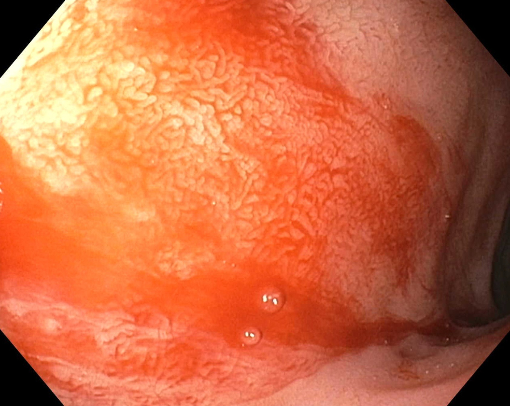
Case Description/Methods: A 28-year-old man with cirrhosis due to alcohol presented to our facility after relapsing from alcohol use disorder and was found to have hematochezia and severe anemia, with Hgb 4.3, Plt 76, INR 3.45. Endoscopy showed two columns of small, non-bleeding esophageal varices, a large amount of fresh red blood in the stomach, and mild portal hypertension gastropathy without varices, ulceration, arteriovenous malformations, or GAVE. Advancement into the duodenum showed a patch at the 9 o’clock position with significant and active bleeding [Figure 1]. Given patients underlying coagulopathy, hemostatic powder was applied liberally with successful hemostasis. Repeat endoscopies two days and nine days later both showed no evidence of further bleeding from the duodenal vascular ectasia, and the patient was discharged. One week later, the patient developed acute hepatic encephalopathy and was admitted. During the admission he subsequently had massive hematochezia with hemodynamic instability. His family elected to transition him to comfort care thus a repeat endoscopy was not performed. Autopsy revealed two liters of fresh blood within the stomach and only a deflated esophageal varix, suggesting that his source of bleed is from the DUVE.
Discussion: Isolated DUVE without evidence of GAVE is exceedingly rare, with only one reported isolated case of DUVE. Here we present a case of isolated DUVE that seemingly caused multiple episodes of severe GI bleeding and ultimately death. Previous case reports demonstrated treatment of ectasias using argon plasma coagulation, but due to this patient’s underlying coagulopathy, only hemostatic powder was applied, resulting in temporary hemostasis. This patient’s presentation and outcome highlights a need for further research in the management of this rare but serious type of gastrointestinal bleeding.

Disclosures:
David Cheung, MD1, Samuel S. Ji, DO2, Vamsi Vemireddy, MD2, Amirali Tavangar, MD2, James Han, MD2, Jason Samarasena, MD, MBA, FACG3. E0325 - Isolated Duodenal Vascular Ectasia Causing Fatal Gastrointestinal Bleeding, ACG 2022 Annual Scientific Meeting Abstracts. Charlotte, NC: American College of Gastroenterology.
1UCI Medical Center, Orange, CA; 2University of California Irvine, Orange, CA; 3UC Irvine, Orange, CA
Introduction: Vascular ectasias are a common cause of acute upper gastrointestinal bleeding due to dilation and fragility of gastrointestinal capillaries. They are most commonly seen as gastric antral vascular ectasia (GAVE), and are associated with conditions like cirrhosis, ESRD, and autoimmune connective tissue disease. Duodenal vascular ectasias (DUVE) are rare and are usually present with concomitant GAVE. Here we present the case of an isolated DUVE that was difficult to manage and ultimately fatal.
Case Description/Methods: A 28-year-old man with cirrhosis due to alcohol presented to our facility after relapsing from alcohol use disorder and was found to have hematochezia and severe anemia, with Hgb 4.3, Plt 76, INR 3.45. Endoscopy showed two columns of small, non-bleeding esophageal varices, a large amount of fresh red blood in the stomach, and mild portal hypertension gastropathy without varices, ulceration, arteriovenous malformations, or GAVE. Advancement into the duodenum showed a patch at the 9 o’clock position with significant and active bleeding [Figure 1]. Given patients underlying coagulopathy, hemostatic powder was applied liberally with successful hemostasis. Repeat endoscopies two days and nine days later both showed no evidence of further bleeding from the duodenal vascular ectasia, and the patient was discharged. One week later, the patient developed acute hepatic encephalopathy and was admitted. During the admission he subsequently had massive hematochezia with hemodynamic instability. His family elected to transition him to comfort care thus a repeat endoscopy was not performed. Autopsy revealed two liters of fresh blood within the stomach and only a deflated esophageal varix, suggesting that his source of bleed is from the DUVE.
Discussion: Isolated DUVE without evidence of GAVE is exceedingly rare, with only one reported isolated case of DUVE. Here we present a case of isolated DUVE that seemingly caused multiple episodes of severe GI bleeding and ultimately death. Previous case reports demonstrated treatment of ectasias using argon plasma coagulation, but due to this patient’s underlying coagulopathy, only hemostatic powder was applied, resulting in temporary hemostasis. This patient’s presentation and outcome highlights a need for further research in the management of this rare but serious type of gastrointestinal bleeding.

Figure: Duodenal vascular ectasia bleeding
Disclosures:
David Cheung indicated no relevant financial relationships.
Samuel Ji indicated no relevant financial relationships.
Vamsi Vemireddy indicated no relevant financial relationships.
Amirali Tavangar indicated no relevant financial relationships.
James Han indicated no relevant financial relationships.
Jason Samarasena: Conmed – Consultant. Docbot – Stock Options. Mauna Kea – Consultant. Olympus – Consultant. Ovesco – Consultant. Steris – Consultant.
David Cheung, MD1, Samuel S. Ji, DO2, Vamsi Vemireddy, MD2, Amirali Tavangar, MD2, James Han, MD2, Jason Samarasena, MD, MBA, FACG3. E0325 - Isolated Duodenal Vascular Ectasia Causing Fatal Gastrointestinal Bleeding, ACG 2022 Annual Scientific Meeting Abstracts. Charlotte, NC: American College of Gastroenterology.
