Back
Poster Session E - Tuesday Afternoon
E0241 - Dysphagia and Esophageal Mass: Cancer or Actinomycosis?
Tuesday, October 25, 2022
3:00 PM – 5:00 PM ET
Location: Crown Ballroom

Tian Li, MD, MS
SUNY Downstate Health Sciences University
Brooklyn, NY
Presenting Author(s)
Tian Li, MD, MS1, Siying Chen, BS2, Kevin Tin, MD3, Snow Trinh Nguyen, MD, MS4
1SUNY Downstate Health Sciences University, Brooklyn, NY; 2University of Buffalo, State University of New York, Brooklyn, NY; 3New York, NY; 4NYU Langone Health, New York, NY
Introduction: Esophageal actinomycosis is a rare type of esophageal infection and presents as erosions or ulcers under endoscopy. Here we present a 63-year-old woman who complains of dysphagia, with biopsy showing actinomyces infection but repeat biopsy revealed squamous cell carcinoma.
Case Description/Methods: A 63-year-old Chinese female with a history of gastritis presented with solid food dysphagia and epigastric pain for over a month. The pain was not improved after proton pump inhibitors but was relieved by self-induced vomiting. Review of systems showed 10lb weight loss over a month with a recent history of self-resolved hematemesis. Prior esophagogastroduodenoscopy (EGD) and colonoscopy were normal 3 years ago.
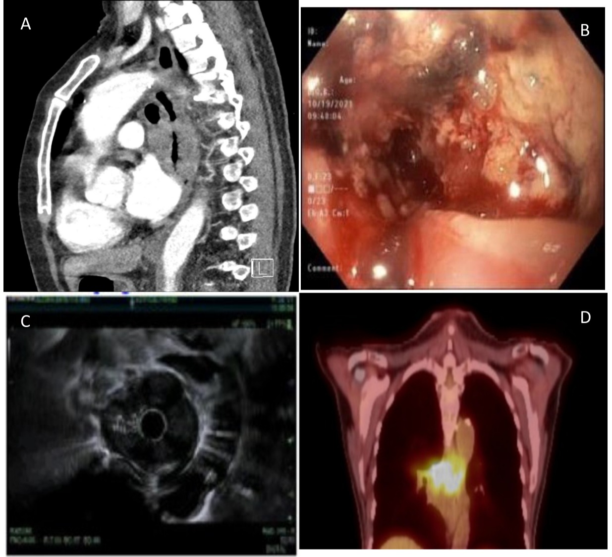
CT of the chest demonstrated a 1.7 cm circumferential mass in the mid-esophagus with luminal narrowing (Figure 1A). EGD discovered a friable soft circumferential mass 26-29 cm from the incisors that are not actively bleeding but covered with blood clots (Figure 1B). Biopsy showed esophageal mucosal ulceration and actinomyces infection. The patient was started on amoxicillin to treat actinomyces infection. Meanwhile, the patient underwent a repeat EGD for rebiopsy given concerns for malignancy, which resulted in poorly differentiated squamous cell carcinoma. Endoscopic ultrasonography (EUS) was performed for staging but was limited staged uT3N1Mx as the mass could not be traversed by echoendoscopy (Figure 1C). Later PET-CT illustrated locally advanced disease with atrium involvement (Figure 1D). The patient underwent neoadjuvant chemotherapy with carboplatin-taxol and radiation followed by esophagogastrectomy for curative intent.
Discussion: Actinomyces are facultatively anaerobic, Gram-negative bacilli. They commensally live within the oral cavity and gastrointestinal tract. Most esophageal actinomyces (EA) infection was previously described to resemble esophagitis or esophageal ulcers, with the endoscopic description being extensive necrotic areas. EA typically presents with dysphagia and odynophagia, particularly in immunocompromised patients or with malignancies. Actinomyces species capitalize on tissue injury or mucosal breach to invade adjacent structures and spreads regardless of anatomical barrier, thus mimicking malignancy. In our case, the local tissue damage caused by neoplastic disease or irradiation predisposed the actinomyces infection. Clinicians need to have a high index of suspicion and clinical knowledge regarding its unusual presentations and ability to mimic malignancy.

Disclosures:
Tian Li, MD, MS1, Siying Chen, BS2, Kevin Tin, MD3, Snow Trinh Nguyen, MD, MS4. E0241 - Dysphagia and Esophageal Mass: Cancer or Actinomycosis?, ACG 2022 Annual Scientific Meeting Abstracts. Charlotte, NC: American College of Gastroenterology.
1SUNY Downstate Health Sciences University, Brooklyn, NY; 2University of Buffalo, State University of New York, Brooklyn, NY; 3New York, NY; 4NYU Langone Health, New York, NY
Introduction: Esophageal actinomycosis is a rare type of esophageal infection and presents as erosions or ulcers under endoscopy. Here we present a 63-year-old woman who complains of dysphagia, with biopsy showing actinomyces infection but repeat biopsy revealed squamous cell carcinoma.
Case Description/Methods: A 63-year-old Chinese female with a history of gastritis presented with solid food dysphagia and epigastric pain for over a month. The pain was not improved after proton pump inhibitors but was relieved by self-induced vomiting. Review of systems showed 10lb weight loss over a month with a recent history of self-resolved hematemesis. Prior esophagogastroduodenoscopy (EGD) and colonoscopy were normal 3 years ago.
CT of the chest demonstrated a 1.7 cm circumferential mass in the mid-esophagus with luminal narrowing (Figure 1A). EGD discovered a friable soft circumferential mass 26-29 cm from the incisors that are not actively bleeding but covered with blood clots (Figure 1B). Biopsy showed esophageal mucosal ulceration and actinomyces infection. The patient was started on amoxicillin to treat actinomyces infection. Meanwhile, the patient underwent a repeat EGD for rebiopsy given concerns for malignancy, which resulted in poorly differentiated squamous cell carcinoma. Endoscopic ultrasonography (EUS) was performed for staging but was limited staged uT3N1Mx as the mass could not be traversed by echoendoscopy (Figure 1C). Later PET-CT illustrated locally advanced disease with atrium involvement (Figure 1D). The patient underwent neoadjuvant chemotherapy with carboplatin-taxol and radiation followed by esophagogastrectomy for curative intent.
Discussion: Actinomyces are facultatively anaerobic, Gram-negative bacilli. They commensally live within the oral cavity and gastrointestinal tract. Most esophageal actinomyces (EA) infection was previously described to resemble esophagitis or esophageal ulcers, with the endoscopic description being extensive necrotic areas. EA typically presents with dysphagia and odynophagia, particularly in immunocompromised patients or with malignancies. Actinomyces species capitalize on tissue injury or mucosal breach to invade adjacent structures and spreads regardless of anatomical barrier, thus mimicking malignancy. In our case, the local tissue damage caused by neoplastic disease or irradiation predisposed the actinomyces infection. Clinicians need to have a high index of suspicion and clinical knowledge regarding its unusual presentations and ability to mimic malignancy.

Figure: Figure1 A. CT of the chest demonstrated a 1.7 cm mass present in the mid-esophagus with lumen narrowing. B. A friable mass without active bleeding but covered with blood clots on endoscopy finding. C. Limited staged uT3N1Mx under EUS D. PET-CT showed locally advanced disease with atrium involvement.
Disclosures:
Tian Li indicated no relevant financial relationships.
Siying Chen indicated no relevant financial relationships.
Kevin Tin indicated no relevant financial relationships.
Snow Trinh Nguyen indicated no relevant financial relationships.
Tian Li, MD, MS1, Siying Chen, BS2, Kevin Tin, MD3, Snow Trinh Nguyen, MD, MS4. E0241 - Dysphagia and Esophageal Mass: Cancer or Actinomycosis?, ACG 2022 Annual Scientific Meeting Abstracts. Charlotte, NC: American College of Gastroenterology.
