Back
Poster Session E - Tuesday Afternoon
E0224 - Esophageal Neuroendocrine Carcinoma Presenting After Definitive Chemoradiation of Squamous Cell Carcinoma in the Same Location
Tuesday, October 25, 2022
3:00 PM – 5:00 PM ET
Location: Crown Ballroom
.jpg)
Zarian Prenatt, DO
St. Luke's University Health Network
Bethlehem, PA
Presenting Author(s)
Zarian Prenatt, DO, Hammad Liaquat, MD, Brittney Shupp, DO, Lisa Stoll, MD, MPH, Yecheskel Schneider, MD
St. Luke's University Health Network, Bethlehem, PA
Introduction: Esophageal neuroendocrine carcinoma (ENEC) is an extremely rare and highly aggressive malignancy that carries a poor prognosis. We present a rare case of ENEC presenting years after chemoradiation of squamous cell carcinoma (SCC) in the same location.
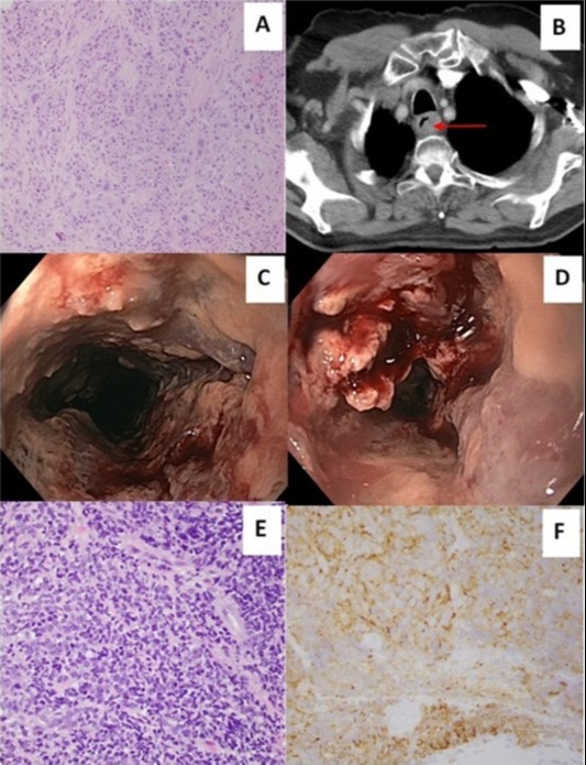
Case Description/Methods: An 85-year-old male presented for oncology follow-up with difficulty swallowing. The patient had a past medical history significant for SCC of the upper esophagus (Image A), for which he received chemoradiation with subsequent 3-year remission confirmed by serial imaging and endoscopy. Given his new-onset dysphagia, a CT of the neck and chest with contrast (Image B) was completed and showed soft tissue thickening in the upper esophagus at the location of prior SCC. The patient underwent EGD, which showed severe friable and ulcerated mucosa in the upper third of the esophagus (Image C & D). Biopsy revealed high-grade malignancy consistent with poorly differentiated neuroendocrine carcinoma, small cell type (Image E). Immunohistochemistry staining was positive for synaptophysin (Image F), pan-CK AE1/3, TTF1, and CD56. The Ki-67 mitotic proliferation index was 80-90%. He underwent chemotherapy with etoposide and carboplatin, but unfortunately had progression of his disease evident on subsequent CT imaging. He was found to have metastatic lesions in the scalp on head CT and passed away in hospice 14 months after diagnosis.
Discussion: To our knowledge, occurrence of ENEC in a site of prior esophageal SCC is a rarity. It could represent a case of radiation-induced malignancy (RIM) based on the temporal relation to his previous SCC treatment. Approximately 8% of second solid cancers may be related to radiation treatment, developing years after initial diagnosis and treatment of the first cancer. Unfortunately, management of ENEC is challenging because treatment strategies have not been well established due to the small number of cases reported in the literature. Patients and providers should discuss the possibility of developing secondary cancer from radiotherapy, and patients who have received radiation should be followed closely for RIM.

Disclosures:
Zarian Prenatt, DO, Hammad Liaquat, MD, Brittney Shupp, DO, Lisa Stoll, MD, MPH, Yecheskel Schneider, MD. E0224 - Esophageal Neuroendocrine Carcinoma Presenting After Definitive Chemoradiation of Squamous Cell Carcinoma in the Same Location, ACG 2022 Annual Scientific Meeting Abstracts. Charlotte, NC: American College of Gastroenterology.
St. Luke's University Health Network, Bethlehem, PA
Introduction: Esophageal neuroendocrine carcinoma (ENEC) is an extremely rare and highly aggressive malignancy that carries a poor prognosis. We present a rare case of ENEC presenting years after chemoradiation of squamous cell carcinoma (SCC) in the same location.
Case Description/Methods: An 85-year-old male presented for oncology follow-up with difficulty swallowing. The patient had a past medical history significant for SCC of the upper esophagus (Image A), for which he received chemoradiation with subsequent 3-year remission confirmed by serial imaging and endoscopy. Given his new-onset dysphagia, a CT of the neck and chest with contrast (Image B) was completed and showed soft tissue thickening in the upper esophagus at the location of prior SCC. The patient underwent EGD, which showed severe friable and ulcerated mucosa in the upper third of the esophagus (Image C & D). Biopsy revealed high-grade malignancy consistent with poorly differentiated neuroendocrine carcinoma, small cell type (Image E). Immunohistochemistry staining was positive for synaptophysin (Image F), pan-CK AE1/3, TTF1, and CD56. The Ki-67 mitotic proliferation index was 80-90%. He underwent chemotherapy with etoposide and carboplatin, but unfortunately had progression of his disease evident on subsequent CT imaging. He was found to have metastatic lesions in the scalp on head CT and passed away in hospice 14 months after diagnosis.
Discussion: To our knowledge, occurrence of ENEC in a site of prior esophageal SCC is a rarity. It could represent a case of radiation-induced malignancy (RIM) based on the temporal relation to his previous SCC treatment. Approximately 8% of second solid cancers may be related to radiation treatment, developing years after initial diagnosis and treatment of the first cancer. Unfortunately, management of ENEC is challenging because treatment strategies have not been well established due to the small number of cases reported in the literature. Patients and providers should discuss the possibility of developing secondary cancer from radiotherapy, and patients who have received radiation should be followed closely for RIM.

Figure: (A) H&E stain showing moderately differentiated SCC. (B) CT chest showing soft tissue thickening (red arrow) in the proximal third of the esophagus compatible with recurrent tumor. (C, D) EGD showing friable, hemorrhagic, and ulcerated mucosa in the upper third of the esophagus. (E) Sheets of neuroendocrine carcinoma showing small blue cells with scant cytoplasm and nuclear molding. (F) Synaptophysin immunohistochemical stain showing diffuse cytoplasmic staining confirming neuroendocrine nature of the tumor.
Disclosures:
Zarian Prenatt indicated no relevant financial relationships.
Hammad Liaquat indicated no relevant financial relationships.
Brittney Shupp indicated no relevant financial relationships.
Lisa Stoll indicated no relevant financial relationships.
Yecheskel Schneider indicated no relevant financial relationships.
Zarian Prenatt, DO, Hammad Liaquat, MD, Brittney Shupp, DO, Lisa Stoll, MD, MPH, Yecheskel Schneider, MD. E0224 - Esophageal Neuroendocrine Carcinoma Presenting After Definitive Chemoradiation of Squamous Cell Carcinoma in the Same Location, ACG 2022 Annual Scientific Meeting Abstracts. Charlotte, NC: American College of Gastroenterology.
