Back
Poster Session E - Tuesday Afternoon
E0226 - Mixed Adenoneuroendocrine Carcinoma at the Gastroesophageal Junction: A Case Report
Tuesday, October 25, 2022
3:00 PM – 5:00 PM ET
Location: Crown Ballroom

Forrest K. Smith, DO
Appalachian Regional Healthcare
Whitesburg, KY
Presenting Author(s)
Joshua K. Jenkins, MS1, Forrest K. Smith, DO2, Sandhya Kolagatla, MD2, Shweta Chaudhary, MD3, Stephen Mularz, DO2
1Lincoln Memorial University-DeBusk College of Osteopathic Medicine, Hazard, KY; 2Appalachian Regional Healthcare, Whitesburg, KY; 3Appalachian Regional Healthcare, Hazard, KY
Introduction: Mixed adenoneuroendocrine carcinomas (MANECs) are a type of mixed neuroendocrine non-neuroendocrine neoplasm (MiNEN), which is a very rare and highly aggressive group of neoplasms found in the gastro-entero-pancreatic (GEP) tract. MANECs comprise both an adenocarcinoma and neuroendocrine carcinoma component. Currently the literature regarding these neoplasms is limited to only retrospective studies and case reports. There have been no prospective randomized trials to support any particular diagnostic algorithm or treatment plan.
Case Description/Methods: A 56-year-old male smoker presented to the emergency room with two weeks of intermittent dysphagia to solids and liquids, epigastric abdominal pain, and melena associated with a 23-pound unintentional weight loss in one month. Computed tomography (CT) revealed a narrowed and thickened distal esophagus. Subsequent esophagoduodenoscopy (EGD) revealed a malignant appearing stricture at the gastroesophageal junction (GEJ) with a large, polypoid mass bulging from the gastric cardia. Histopathological analysis revealed poorly differentiated adenocarcinoma admixed with high-grade (Grade 3) neuroendocrine differentiation. Immunohistochemistry was significant for AE1/AE3, CK7, CDX2, CK20, and synaptophysin positivity. The patient was referred to an external facility for neoadjuvant chemoradiation and local surgical resection via Ivor-Lewis esophagectomy.
Discussion: MANECs are rare tumors of the alimentary tract, composed of neuroendocrine and non-neuroendocrine carcinoma components. These tumors are highly aggressive and often fatal. We found nine other cases of MANEC discovered at the GEJ in the English medical literature. Given this rare occurrence, we aim to add to the existing literature with another case to support diagnostic and treatment modalities for these highly aggressive mixed neoplasms in the future. This case is currently ongoing and we are following closely as the patient receives further treatment.

Disclosures:
Joshua K. Jenkins, MS1, Forrest K. Smith, DO2, Sandhya Kolagatla, MD2, Shweta Chaudhary, MD3, Stephen Mularz, DO2. E0226 - Mixed Adenoneuroendocrine Carcinoma at the Gastroesophageal Junction: A Case Report, ACG 2022 Annual Scientific Meeting Abstracts. Charlotte, NC: American College of Gastroenterology.
1Lincoln Memorial University-DeBusk College of Osteopathic Medicine, Hazard, KY; 2Appalachian Regional Healthcare, Whitesburg, KY; 3Appalachian Regional Healthcare, Hazard, KY
Introduction: Mixed adenoneuroendocrine carcinomas (MANECs) are a type of mixed neuroendocrine non-neuroendocrine neoplasm (MiNEN), which is a very rare and highly aggressive group of neoplasms found in the gastro-entero-pancreatic (GEP) tract. MANECs comprise both an adenocarcinoma and neuroendocrine carcinoma component. Currently the literature regarding these neoplasms is limited to only retrospective studies and case reports. There have been no prospective randomized trials to support any particular diagnostic algorithm or treatment plan.
Case Description/Methods: A 56-year-old male smoker presented to the emergency room with two weeks of intermittent dysphagia to solids and liquids, epigastric abdominal pain, and melena associated with a 23-pound unintentional weight loss in one month. Computed tomography (CT) revealed a narrowed and thickened distal esophagus. Subsequent esophagoduodenoscopy (EGD) revealed a malignant appearing stricture at the gastroesophageal junction (GEJ) with a large, polypoid mass bulging from the gastric cardia. Histopathological analysis revealed poorly differentiated adenocarcinoma admixed with high-grade (Grade 3) neuroendocrine differentiation. Immunohistochemistry was significant for AE1/AE3, CK7, CDX2, CK20, and synaptophysin positivity. The patient was referred to an external facility for neoadjuvant chemoradiation and local surgical resection via Ivor-Lewis esophagectomy.
Discussion: MANECs are rare tumors of the alimentary tract, composed of neuroendocrine and non-neuroendocrine carcinoma components. These tumors are highly aggressive and often fatal. We found nine other cases of MANEC discovered at the GEJ in the English medical literature. Given this rare occurrence, we aim to add to the existing literature with another case to support diagnostic and treatment modalities for these highly aggressive mixed neoplasms in the future. This case is currently ongoing and we are following closely as the patient receives further treatment.

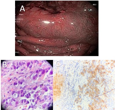
Figure: Figure 1: A) Endoscopic image demonstrating polypoid mass protruding from gastroesophageal junction into gastric cardia under narrow band imaging technology. B) ) H&E stain of the specimen demonstrating poorly differentiated adenocarcinoma with signet ring features, x400. C) Immunohistochemical staining demonstrating positivity for CK7 in the neuroendocrine carcinoma component (x100)
Disclosures:
Joshua Jenkins indicated no relevant financial relationships.
Forrest Smith indicated no relevant financial relationships.
Sandhya Kolagatla indicated no relevant financial relationships.
Shweta Chaudhary indicated no relevant financial relationships.
Stephen Mularz indicated no relevant financial relationships.
Joshua K. Jenkins, MS1, Forrest K. Smith, DO2, Sandhya Kolagatla, MD2, Shweta Chaudhary, MD3, Stephen Mularz, DO2. E0226 - Mixed Adenoneuroendocrine Carcinoma at the Gastroesophageal Junction: A Case Report, ACG 2022 Annual Scientific Meeting Abstracts. Charlotte, NC: American College of Gastroenterology.
