Back
Poster Session E - Tuesday Afternoon
E0116 - Now You See Me, Now You Don’t: A Case of Campylobacter Jejuni Colitis Presenting as a Colonic Mass
Tuesday, October 25, 2022
3:00 PM – 5:00 PM ET
Location: Crown Ballroom

Randy Leibowitz, DO
Mount Sinai St. Luke's and Mount Sinai Roosevelt
New York, NY
Presenting Author(s)
Randy Leibowitz, DO1, Frederick Rozenshteyn, MD2, Samuel Daniel, MD3
1Mount Sinai St. Luke's and Mount Sinai Roosevelt, New York, NY; 2Icahn School of Medicine at Mount Sinai Morningside-West, New York, NY; 3Mount Sinai Beth Israel, Morningside and West, New York, NY
Introduction: Campylobacter Jejuni (C. Jejuni) is responsible for 3-6% of diarrheal illnesses in the United States. Inoculation follows ingestion of a contaminated source such as poultry, unpasteurized milk, or drinking water. The presentation of a C. Jejuni infection may radiographically and endoscopically mimic other pathologies such as colonic malignancy and inflammatory bowel disease (IBD).
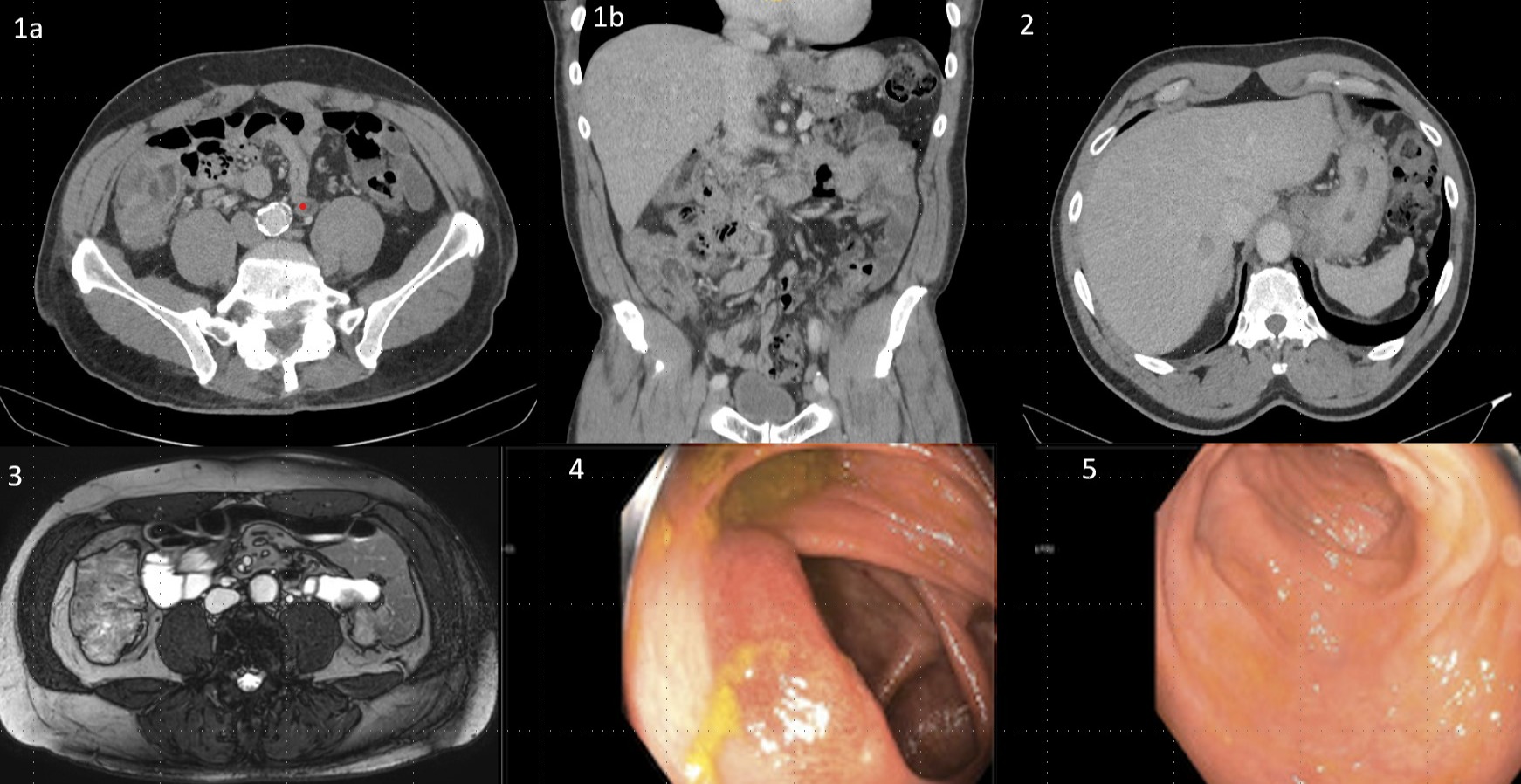
Case Description/Methods: A 66-year old man with a past medical history of prostate cancer in remission and a 6mm colonic polyp removed 5 years prior presented to the emergency department with 5 days of watery diarrhea. He complained of 6 watery bowel movements per day associated with colicky abdominal pain and bloating. He was hemodynamically stable with a physical exam notable for severe tenderness in the right lower quadrant. Serum chemistries did not reveal any abnormalities. Computed tomography revealed severe, focal bowel wall thickening in the ascending colon and a 1.7cm hypodense lesion in the right hepatic lobe which was confirmed on magnetic resonance of the abdomen. The radiologist’s interpretation emphasized concern for colonic malignancy with metastasis to the liver.
Endoscopic evaluation revealed diffusely erythematous, granular and ulcerated mucosa throughout the colon with concern for infectious vs inflammatory colitis. Histologic evaluation of ascending colonic mucosal biopsy revealed moderately active chronic colitis with extensive cryptitis and crypt abscesses. Stool polymerase chain reaction was notable for C. Jejuni infection and the patient achieved clinical and endoscopic remission without antimicrobial therapy.
Discussion: In patients with C. Jejuni enterocolitis, radiographic imaging may reveal signs of bowel wall edema or ulcerations which may resemble colonic malignancy or IBD. Endoscopic findings of campylobacter enterocolitis are non-specific, however, may reveal edematous, erythematous, and friable mucosa which may be associated with hemorrhage. Inflamed mucosa can be either isolated or discontinuous. Colonic ulcers may present as either aphthous or linear (may resemble cobblestoning as seen in Crohn's Disease). Histologic evaluation can reveal cryptitis and crypt abscess formation similar to findings of ulcerative colitis.
This case highlights the range of radiographic and endoscopic presentations of C. Jejuni Colitis. Infectious colitis should remain on the differential for radiographic evidence of colonic edema in the right clinical setting.

Disclosures:
Randy Leibowitz, DO1, Frederick Rozenshteyn, MD2, Samuel Daniel, MD3. E0116 - Now You See Me, Now You Don’t: A Case of Campylobacter Jejuni Colitis Presenting as a Colonic Mass, ACG 2022 Annual Scientific Meeting Abstracts. Charlotte, NC: American College of Gastroenterology.
1Mount Sinai St. Luke's and Mount Sinai Roosevelt, New York, NY; 2Icahn School of Medicine at Mount Sinai Morningside-West, New York, NY; 3Mount Sinai Beth Israel, Morningside and West, New York, NY
Introduction: Campylobacter Jejuni (C. Jejuni) is responsible for 3-6% of diarrheal illnesses in the United States. Inoculation follows ingestion of a contaminated source such as poultry, unpasteurized milk, or drinking water. The presentation of a C. Jejuni infection may radiographically and endoscopically mimic other pathologies such as colonic malignancy and inflammatory bowel disease (IBD).
Case Description/Methods: A 66-year old man with a past medical history of prostate cancer in remission and a 6mm colonic polyp removed 5 years prior presented to the emergency department with 5 days of watery diarrhea. He complained of 6 watery bowel movements per day associated with colicky abdominal pain and bloating. He was hemodynamically stable with a physical exam notable for severe tenderness in the right lower quadrant. Serum chemistries did not reveal any abnormalities. Computed tomography revealed severe, focal bowel wall thickening in the ascending colon and a 1.7cm hypodense lesion in the right hepatic lobe which was confirmed on magnetic resonance of the abdomen. The radiologist’s interpretation emphasized concern for colonic malignancy with metastasis to the liver.
Endoscopic evaluation revealed diffusely erythematous, granular and ulcerated mucosa throughout the colon with concern for infectious vs inflammatory colitis. Histologic evaluation of ascending colonic mucosal biopsy revealed moderately active chronic colitis with extensive cryptitis and crypt abscesses. Stool polymerase chain reaction was notable for C. Jejuni infection and the patient achieved clinical and endoscopic remission without antimicrobial therapy.
Discussion: In patients with C. Jejuni enterocolitis, radiographic imaging may reveal signs of bowel wall edema or ulcerations which may resemble colonic malignancy or IBD. Endoscopic findings of campylobacter enterocolitis are non-specific, however, may reveal edematous, erythematous, and friable mucosa which may be associated with hemorrhage. Inflamed mucosa can be either isolated or discontinuous. Colonic ulcers may present as either aphthous or linear (may resemble cobblestoning as seen in Crohn's Disease). Histologic evaluation can reveal cryptitis and crypt abscess formation similar to findings of ulcerative colitis.
This case highlights the range of radiographic and endoscopic presentations of C. Jejuni Colitis. Infectious colitis should remain on the differential for radiographic evidence of colonic edema in the right clinical setting.

Figure: Figure 1a,1b: CT Abdomen/Pelvis showing ascending colon wall thickening resembling mass; Figure 2: CT of Hypoechoic liver mass; Figure 3: T2-weighted MRI Abdomen of ascending colon wall thickening resembling a mass; Figure 4: Colonoscopy image revealing inflamed mucosa of ascending colon; Figure 5: Ulcerated mucosa of the sigmoid colon
Disclosures:
Randy Leibowitz indicated no relevant financial relationships.
Frederick Rozenshteyn indicated no relevant financial relationships.
Samuel Daniel indicated no relevant financial relationships.
Randy Leibowitz, DO1, Frederick Rozenshteyn, MD2, Samuel Daniel, MD3. E0116 - Now You See Me, Now You Don’t: A Case of Campylobacter Jejuni Colitis Presenting as a Colonic Mass, ACG 2022 Annual Scientific Meeting Abstracts. Charlotte, NC: American College of Gastroenterology.
