Back
Poster Session E - Tuesday Afternoon
E0110 - Chilaiditi’s Sign
Tuesday, October 25, 2022
3:00 PM – 5:00 PM ET
Location: Crown Ballroom
.jpg)
Brian M. Fung, MD
University of Arizona College of Medicine
Phoenix, AZ
Presenting Author(s)
Brian M. Fung, MD1, Jen-Jung Pan, MD, PhD1, Jen-Tzer Gau, MD, PhD2
1University of Arizona College of Medicine, Phoenix, AZ; 2Ohio University Heritage College of Osteopathic Medicine, Athens, OH
Introduction: Chilaiditi’s sign refers to the incidental radiographic finding of large bowel interposed between the diaphragm and the liver. This finding can frequently be mistaken as pneumoperitoneum, a finding that is often thought of as a surgical emergency. In the following case, we describe a patient who was found to have Chilaiditi’s sign on computed tomography (CT) imaging.
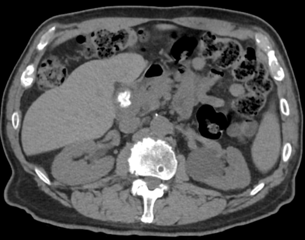
Case Description/Methods: A 75-year-old male with history of bladder cancer status post resection six days prior presented to the emergency room for lower abdominal pain and hematuria. He denied having any nausea, vomiting, or changes in bowel habits. Except for a hemoglobin of 12.6 g/dL, basic laboratory testing including a complete blood count and comprehensive metabolic panel was within normal ranges. Physical examination revealed suprapubic tenderness but was otherwise unremarkable. To further evaluate his symptoms, a CT of the abdomen and pelvis was performed, revealing a hematoma in the bladder wall and the appearance of free air under the diaphragm. However, upon further inspection, the air was noted to be within the lumen of a segment of colon which was interposed between the liver and the diaphragm (Figure 1), consistent with Chilaiditi’s sign. This was thought to be an incidental finding, and the patient was referred to urology for further management of his bladder cancer.
Discussion: Chilaiditi’s sign, or hepatodiaphragmatic interposition of the colon, is a rare radiological finding with a reported incidence of 0.025% to 0.28%. Due to air frequently found in the colon, patients with this condition can appear to have pneumoperitoneum due to the colon being near the diaphragm in an abnormal location. The finding is more common in males, older patients, and patients with dolichocolon, chronic lung disease, liver disease, or ascites. Although the cause of this condition is not clearly understood, absence or laxity of the ligament supporting the transverse colon or the falciform ligament is thought to contribute to this condition. Patients with this finding are generally asymptomatic and do not require any treatment. In rare cases, the finding is associated with clinical symptoms, such as abdominal pain, nausea, vomiting, and constipation; in these circumstances, the condition is termed Chilaiditi’s syndrome.

Disclosures:
Brian M. Fung, MD1, Jen-Jung Pan, MD, PhD1, Jen-Tzer Gau, MD, PhD2. E0110 - Chilaiditi’s Sign, ACG 2022 Annual Scientific Meeting Abstracts. Charlotte, NC: American College of Gastroenterology.
1University of Arizona College of Medicine, Phoenix, AZ; 2Ohio University Heritage College of Osteopathic Medicine, Athens, OH
Introduction: Chilaiditi’s sign refers to the incidental radiographic finding of large bowel interposed between the diaphragm and the liver. This finding can frequently be mistaken as pneumoperitoneum, a finding that is often thought of as a surgical emergency. In the following case, we describe a patient who was found to have Chilaiditi’s sign on computed tomography (CT) imaging.
Case Description/Methods: A 75-year-old male with history of bladder cancer status post resection six days prior presented to the emergency room for lower abdominal pain and hematuria. He denied having any nausea, vomiting, or changes in bowel habits. Except for a hemoglobin of 12.6 g/dL, basic laboratory testing including a complete blood count and comprehensive metabolic panel was within normal ranges. Physical examination revealed suprapubic tenderness but was otherwise unremarkable. To further evaluate his symptoms, a CT of the abdomen and pelvis was performed, revealing a hematoma in the bladder wall and the appearance of free air under the diaphragm. However, upon further inspection, the air was noted to be within the lumen of a segment of colon which was interposed between the liver and the diaphragm (Figure 1), consistent with Chilaiditi’s sign. This was thought to be an incidental finding, and the patient was referred to urology for further management of his bladder cancer.
Discussion: Chilaiditi’s sign, or hepatodiaphragmatic interposition of the colon, is a rare radiological finding with a reported incidence of 0.025% to 0.28%. Due to air frequently found in the colon, patients with this condition can appear to have pneumoperitoneum due to the colon being near the diaphragm in an abnormal location. The finding is more common in males, older patients, and patients with dolichocolon, chronic lung disease, liver disease, or ascites. Although the cause of this condition is not clearly understood, absence or laxity of the ligament supporting the transverse colon or the falciform ligament is thought to contribute to this condition. Patients with this finding are generally asymptomatic and do not require any treatment. In rare cases, the finding is associated with clinical symptoms, such as abdominal pain, nausea, vomiting, and constipation; in these circumstances, the condition is termed Chilaiditi’s syndrome.

Figure: Interposition of a stool-filled transverse colon between the liver and right hemidiaphragm.
Disclosures:
Brian Fung indicated no relevant financial relationships.
Jen-Jung Pan indicated no relevant financial relationships.
Jen-Tzer Gau indicated no relevant financial relationships.
Brian M. Fung, MD1, Jen-Jung Pan, MD, PhD1, Jen-Tzer Gau, MD, PhD2. E0110 - Chilaiditi’s Sign, ACG 2022 Annual Scientific Meeting Abstracts. Charlotte, NC: American College of Gastroenterology.
