Back
Poster Session D - Tuesday Morning
D0154 - Diagnostic Significance of Rare Colorectal Perineurioma Found on Colonoscopy: A Case Report
Tuesday, October 25, 2022
10:00 AM – 12:00 PM ET
Location: Crown Ballroom

Blaine Massey, DO
Kirk Kerkorian School of Medicine at UNLV
Las Vegas, NV
Presenting Author(s)
Sami Mesgun, BS1, Blaine Massey, DO1, Jose Aponte-Pieras, MD1, Joseph Fayad, MD2
1Kirk Kerkorian School of Medicine at UNLV, Las Vegas, NV; 2VA Southern Nevada Health Care System, Las Vegas, NV
Introduction: Perineuriomas are benign spindle cell neoplasms of the peripheral nerve sheath which seldomly involve the GI tract. Colorectal perineuriomas have an incidence of 0.1%-0.46% of all colonic polyps, usually occurring in the sigmoid colon and rectum, and are often diagnosed incidentally on routine screening colonoscopy. They are not associated with neurofibromatosis syndromes (NF1-2) and require no additional followup. Herein, we describe a case of colonic mucosal perineurioma in a patient referred for colonoscopy after a positive gFOBT.
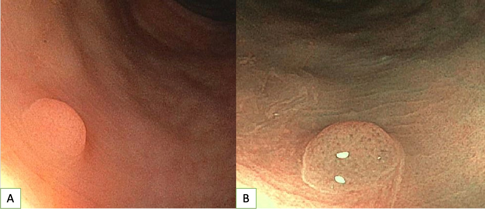
Case Description/Methods: A 57-year-old male with hypertension and dyslipidemia presented to the GI clinic after a positive gFOBT. He was asymptomatic and physical examination was unremarkable. Laboratory evaluation showed mild anemia with Hgb of 13.5 g/dL and a low-normal MCV of 80.1 μm3. Iron studies were normal. Colonoscopy revealed a 2-mm sessile rectosigmoid polyp (Figure 1), which underwent cold snare polypectomy with histopathology notable for bland spindle cells with small elongated nuclei and imperceptible cell borders. No significant nuclear atypia or mitotic activity was identified. Immunohistochemistry (IHC) showed focal epithelial membrane antigen (EMA) staining of stromal cells; S100 stain was negative. These findings were consistent with perineurioma. Remainder of colonoscopy only showed multiple subcentimeter tubulovillous and tubular adenomas of the right colon.
Discussion: Colorectal perineuriomas typically appear as small, solitary sessile polyps less than 6 mm in diameter (median 4 mm). Histologically, they appear as uniformly elongated spindle cells with rare mitotic activity. IHC shows diffuse, strongly positive staining of spindle cells with GLUT1 and claudin 1 and focal or faintly positive EMA staining. Two of the three positive stains generally support the diagnosis. Colorectal perineuriomas lack S100 protein expression unlike other soft tissue neuromas such as schwannomas and neurofibromas. This case highlights the importance of correct diagnosis in order to avoid overtreatment, as these may resemble malignant neoplasms such as gastrointestinal stromal tumors which are histologically similar but stain positive for c-kit/CD117 and DOG-1. These tumors are more common in females, with a median age of 51. They do not recur after excision, and given their benign nature, do not require surveillance after polypectomy.

Disclosures:
Sami Mesgun, BS1, Blaine Massey, DO1, Jose Aponte-Pieras, MD1, Joseph Fayad, MD2. D0154 - Diagnostic Significance of Rare Colorectal Perineurioma Found on Colonoscopy: A Case Report, ACG 2022 Annual Scientific Meeting Abstracts. Charlotte, NC: American College of Gastroenterology.
1Kirk Kerkorian School of Medicine at UNLV, Las Vegas, NV; 2VA Southern Nevada Health Care System, Las Vegas, NV
Introduction: Perineuriomas are benign spindle cell neoplasms of the peripheral nerve sheath which seldomly involve the GI tract. Colorectal perineuriomas have an incidence of 0.1%-0.46% of all colonic polyps, usually occurring in the sigmoid colon and rectum, and are often diagnosed incidentally on routine screening colonoscopy. They are not associated with neurofibromatosis syndromes (NF1-2) and require no additional followup. Herein, we describe a case of colonic mucosal perineurioma in a patient referred for colonoscopy after a positive gFOBT.
Case Description/Methods: A 57-year-old male with hypertension and dyslipidemia presented to the GI clinic after a positive gFOBT. He was asymptomatic and physical examination was unremarkable. Laboratory evaluation showed mild anemia with Hgb of 13.5 g/dL and a low-normal MCV of 80.1 μm3. Iron studies were normal. Colonoscopy revealed a 2-mm sessile rectosigmoid polyp (Figure 1), which underwent cold snare polypectomy with histopathology notable for bland spindle cells with small elongated nuclei and imperceptible cell borders. No significant nuclear atypia or mitotic activity was identified. Immunohistochemistry (IHC) showed focal epithelial membrane antigen (EMA) staining of stromal cells; S100 stain was negative. These findings were consistent with perineurioma. Remainder of colonoscopy only showed multiple subcentimeter tubulovillous and tubular adenomas of the right colon.
Discussion: Colorectal perineuriomas typically appear as small, solitary sessile polyps less than 6 mm in diameter (median 4 mm). Histologically, they appear as uniformly elongated spindle cells with rare mitotic activity. IHC shows diffuse, strongly positive staining of spindle cells with GLUT1 and claudin 1 and focal or faintly positive EMA staining. Two of the three positive stains generally support the diagnosis. Colorectal perineuriomas lack S100 protein expression unlike other soft tissue neuromas such as schwannomas and neurofibromas. This case highlights the importance of correct diagnosis in order to avoid overtreatment, as these may resemble malignant neoplasms such as gastrointestinal stromal tumors which are histologically similar but stain positive for c-kit/CD117 and DOG-1. These tumors are more common in females, with a median age of 51. They do not recur after excision, and given their benign nature, do not require surveillance after polypectomy.

Figure: Figure 1
A. Colonoscopy showing a 2-mm sessile rectosigmoid polyp, confirmed as a perineurioma on pathology
B. Closer image of the same perineurioma, visualized using Narrow Band Imaging (NBI)
A. Colonoscopy showing a 2-mm sessile rectosigmoid polyp, confirmed as a perineurioma on pathology
B. Closer image of the same perineurioma, visualized using Narrow Band Imaging (NBI)
Disclosures:
Sami Mesgun indicated no relevant financial relationships.
Blaine Massey indicated no relevant financial relationships.
Jose Aponte-Pieras indicated no relevant financial relationships.
Joseph Fayad indicated no relevant financial relationships.
Sami Mesgun, BS1, Blaine Massey, DO1, Jose Aponte-Pieras, MD1, Joseph Fayad, MD2. D0154 - Diagnostic Significance of Rare Colorectal Perineurioma Found on Colonoscopy: A Case Report, ACG 2022 Annual Scientific Meeting Abstracts. Charlotte, NC: American College of Gastroenterology.
