Back
Poster Session C - Monday Afternoon
C0064 - Giant Mucinous Cystic Neoplasm of the Pancreas in a 17-Year-Old: A Case Report
Monday, October 24, 2022
3:00 PM – 5:00 PM ET
Location: Crown Ballroom
- GR
Grigoriy Rapoport, MD
UTRGV-DHR Gastroenterology Fellowship Program
Plano, TX
Presenting Author(s)
Grigoriy Rapoport, MD1, Ans Albustamy, MD2, Mohammad Shakhatreh, MD3, Henry Herrera, MD4, Asif Zamir, MD, FACG2
1UTRGV-DHR Gastroenterology Fellowship Program, Plano, TX; 2University of Texas Rio Grande Valley at Doctors Hospital at Renaissance, Edinburg, TX; 3UTRGV-DHR Gastroenterology Fellowship Program, Edinburg, TX; 4Renaissance Gastroenterology at Doctor's Hospital at Renaissance, Edinburg, TX
Introduction: Mucinous cystic neoplasms (MCNs) of the pancreas represent one of the most common primary pancreatic cystic neoplasms, accounting for approximately half of all cases. The probability of pancreatic cystic neoplasms being detected is raising year by year, although they are usually detected between ages 40-60, affecting women more than men. We present an unusual case of a gigantic MCN occurring in a 17-year-old patient.
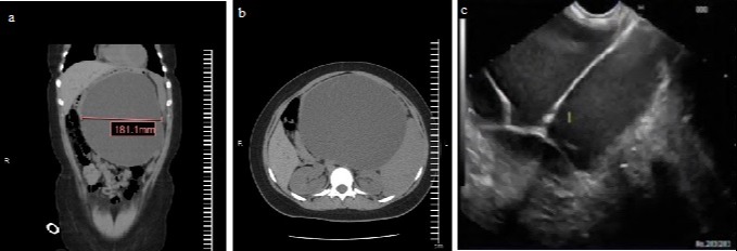
Case Description/Methods: A 17-year-old female with no past medical history presented to the ER with 3-month history of progressive abdominal distention, pain and an unintentional 9lb weight loss for a month. She was hemodynamically compensated, with physical exam significant for epigastric tenderness. Hemoglobin/hematocrit was noted at 6.2 g/dL and 24%, lipase 221 U/L. CT abdomen and pelvis without contrast revealed a large cystic mass in the pancreas, with marked splenomegaly. MRI of the abdomen w/wo contrast confirmed the presence of a large, complex, cystic structure with septations measuring 18x17cm. EUS with FNA of the mass resulted in aspiration of 100 cc of clear, mucoid fluid. Analysis of the aspirate revealed amylase of 8 U/L, glucose 27 mg/dL, and a CA19-9 level of 25,960 IU, raising concern for mucinous cystic neoplasm. She successfully underwent open resection of the mass which measured 20cm, with distal pancreatectomy and splenectomy. Pathology of the mass revealed a mucinous cystic neoplasm with low-grade dysplasia. The patient recovered and was discharged home. She reported significant improvement at a follow up in clinic 2 months later.
Discussion: Giant MCN of the pancreas is described in the literature but has not been observed at such a young age. The incidence of detection of pancreatic cystic lesions increases year by year and is thought to be due to better imaging modalities detecting incidental lesions. To our knowledge, this is the youngest reported patient with symptomatic MCN.

Disclosures:
Grigoriy Rapoport, MD1, Ans Albustamy, MD2, Mohammad Shakhatreh, MD3, Henry Herrera, MD4, Asif Zamir, MD, FACG2. C0064 - Giant Mucinous Cystic Neoplasm of the Pancreas in a 17-Year-Old: A Case Report, ACG 2022 Annual Scientific Meeting Abstracts. Charlotte, NC: American College of Gastroenterology.
1UTRGV-DHR Gastroenterology Fellowship Program, Plano, TX; 2University of Texas Rio Grande Valley at Doctors Hospital at Renaissance, Edinburg, TX; 3UTRGV-DHR Gastroenterology Fellowship Program, Edinburg, TX; 4Renaissance Gastroenterology at Doctor's Hospital at Renaissance, Edinburg, TX
Introduction: Mucinous cystic neoplasms (MCNs) of the pancreas represent one of the most common primary pancreatic cystic neoplasms, accounting for approximately half of all cases. The probability of pancreatic cystic neoplasms being detected is raising year by year, although they are usually detected between ages 40-60, affecting women more than men. We present an unusual case of a gigantic MCN occurring in a 17-year-old patient.
Case Description/Methods: A 17-year-old female with no past medical history presented to the ER with 3-month history of progressive abdominal distention, pain and an unintentional 9lb weight loss for a month. She was hemodynamically compensated, with physical exam significant for epigastric tenderness. Hemoglobin/hematocrit was noted at 6.2 g/dL and 24%, lipase 221 U/L. CT abdomen and pelvis without contrast revealed a large cystic mass in the pancreas, with marked splenomegaly. MRI of the abdomen w/wo contrast confirmed the presence of a large, complex, cystic structure with septations measuring 18x17cm. EUS with FNA of the mass resulted in aspiration of 100 cc of clear, mucoid fluid. Analysis of the aspirate revealed amylase of 8 U/L, glucose 27 mg/dL, and a CA19-9 level of 25,960 IU, raising concern for mucinous cystic neoplasm. She successfully underwent open resection of the mass which measured 20cm, with distal pancreatectomy and splenectomy. Pathology of the mass revealed a mucinous cystic neoplasm with low-grade dysplasia. The patient recovered and was discharged home. She reported significant improvement at a follow up in clinic 2 months later.
Discussion: Giant MCN of the pancreas is described in the literature but has not been observed at such a young age. The incidence of detection of pancreatic cystic lesions increases year by year and is thought to be due to better imaging modalities detecting incidental lesions. To our knowledge, this is the youngest reported patient with symptomatic MCN.

Figure: a) coronal view on CT scan of the mass, b) axial view on CT scan of the mass, c) EUS demonstrating multiple septations of the giant mass
Disclosures:
Grigoriy Rapoport indicated no relevant financial relationships.
Ans Albustamy indicated no relevant financial relationships.
Mohammad Shakhatreh indicated no relevant financial relationships.
Henry Herrera indicated no relevant financial relationships.
Asif Zamir indicated no relevant financial relationships.
Grigoriy Rapoport, MD1, Ans Albustamy, MD2, Mohammad Shakhatreh, MD3, Henry Herrera, MD4, Asif Zamir, MD, FACG2. C0064 - Giant Mucinous Cystic Neoplasm of the Pancreas in a 17-Year-Old: A Case Report, ACG 2022 Annual Scientific Meeting Abstracts. Charlotte, NC: American College of Gastroenterology.
